User login
“Don’t Take Shortcuts,” Endoscopy Researcher Advises
But the work he’s most proud of took place when he was a graduate student at Harvard, working on a master’s degree in epidemiology and biostatistics.
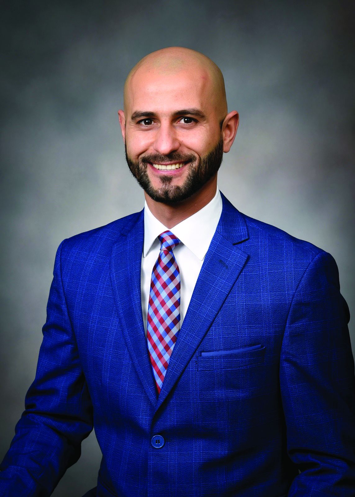
Jovani compared two different types of needles for tissue acquisition with endoscopic ultrasound. His finding that fine needle biopsy is better than fine needle aspiration for lesions isn’t groundbreaking, yet “the reason why I feel proud of that one is because it’s the first paper I did completely by myself,” said Jovani, medical director for advanced therapeutic endoscopy with Gastro Health Florida, in Miami, Florida.
Dr. Jovani has since contributed to countless peer-reviewed articles and book chapters and has presented research findings at meetings across the globe. He will be program director of the upcoming gastroenterology fellowship program at Florida International University School of Medicine, Miami, and participates in several endoscopy panels in the U.S. and in Europe to set guidelines and improve the quality of endoscopic procedures.
Therapeutic endoscopy is a clinical interest of his, specifically in the areas of third space, biliopancreatic and bariatric endoscopy. In an interview, he discussed how he used third space endoscopy to save a patient and improve her quality of life
Indeed, helping patients feel better is the most satisfying part of his career.
“A lot of people may have acute pain or an early cancer or many other problems that they need solving. As a physician, you can be the one who solves it,” said Jovani.
But training in medicine involves hard work, he advised. In the interview, he explained why young doctors should never rely on shortcuts to solve problems.
Therapeutic endoscopy is a specific interest of yours. How has this field advanced since you’ve been practicing gastroenterology?
Dr. Jovani: In the last 10 to 15 years, significant improvements have come along. As an example, lumen-apposing metal stents have revolutionized the way we do therapeutic endoscopy. A lot of procedures were not possible beforehand and we would have to send patients to surgery. Now, these can be done with endoscopy.
Examples include drainage of pancreatic collections, gallbladder drainage, or gastrojejunostomy (a connection between the stomach and the intestine) or reversal of Roux-en-Y gastric bypass to reach and drain the bile duct. Many of these procedures can be done with these metal stents that were not possible beforehand. Bariatric endoscopy is a relatively new field, and that has significantly changed the management of obesity.
There’s also third space endoscopy for the treatment of gastroparesis, achalasia, and early cancer.

What is third space endoscopy and how are you applying it in your practice?
Dr. Jovani: Third space endoscopy refers to a new space that’s created between the mucosa and the muscularis propria into the submucosa. We go in the submucosa, we inject some fluid there, and we cut the submucosa and we separate the mucosa from the muscle.
This allows us to do a lot of procedures. For patients with achalasia, we can tunnel through the submucosa, get into the muscle and perform myotomy, meaning that we can cut the muscle. By doing so, we can treat achalasia with a minimally invasive method. Patients can either go home the next day or even on the same day. The same thing applies for gastroparesis. With early cancer, we can go through in the submucosa, and if the cancer is in the mucosa only, or if it is in the very superficial submucosa, we can treat it without a need for surgery. Sometimes the procedure is simple, but other times it can be very challenging.
Can you discuss a challenging case where you applied third space endoscopy?
Dr. Jovani: It was a gastric cancer case. I did an endoscopic ultrasound for staging purposes. When I saw the lesion, it looked very superficial, like an early cancer of the stomach. I called the surgeon and said I could take it out with endoscopy. And it was in a very difficult location, so it was a very challenging procedure. It took about 12 hours to do it, but I was able to completely take it out. More than a year later, the patient was cancer free and more importantly, we preserved the stomach. Before I did this, she was prepared to undergo total gastrectomy, which meant I would have taken out her entire stomach.
Instead, with this minimally invasive procedure, I was able to take the cancer away and keep the stomach, which preserved her quality of life as well.
When you don’t have the stomach, obviously you adapt, but the quality of life is never the same. The type of food you eat, the frequency of eating, the quality of food you eat is not the same. The fact that we could avoid that in this patient feels very good.
What advice would you give to aspiring medical students?
Dr. Jovani: Do the hard work that’s required to be a doctor. Being a physician is a hard job, but it’s very rewarding. It’s like going to the gym—there really are no shortcuts. You have to do the work, you have to get tired, you have to study hard. You may study things you might not think will be useful to you necessarily in the future field that you choose. If it is GI, you still need to study all the other fields because sometimes patients may have GI diseases that are connecting with other diseases and you won’t know that if you haven’t studied the other diseases.
Patients are not only one disease, but they are also complex patients. Sometimes if you try to correct one disease, you create a complication with the other disease and you might not be aware of that.
Don’t create shortcuts like ChatGPT, things that are becoming fashionable with younger people today. Do the hard work the old way in which you have to memorize things. Knowledge is the only thing that really can help the patient.
Go to GI meetings. Offer to meet people, collaborate, network. Don’t be shy about it. Even if it is not natural to you, just do it. It’ll become more natural as you do it. GI, like any other field, any other endeavor in human society, is something that also depends on interactions. Therefore, it’s good to learn how to interact, how to network, how to do research projects. Even with people from far away, communication is very easy. You don’t really need to do research projects only with people in your local environment. You can do research projects with people who are on the other side of the state or even on the other side of the world.

You place an emphasis on individualized patient care. Can you discuss what that means to you?
Dr. Jovani: It basically means that there isn’t one size fits all in the management of diseases. Obviously there are some general principles that are applicable to everybody, but sometimes for the single specific patient, what works for one patient might not necessarily work for the next patient.
With Endoscopic Retrograde Cholangiopancreatography (ERCP) for example, there are so many things that go into that. Most papilla are in a certain position and it’s relatively easy to cannulate. But there are others that are in very different positions or in different angulations and they might require specific techniques that are not applicable in the majority of cases. You have to adapt to the single patient.How you speak to the patient is also important. Some may prefer a certain type of communication and other patients may prefer another type of communication involving patients or family. You have to adapt to the single patient. You have to understand the different types of personalities and adapt how you explain things or how you communicate disease, or management of disease or even complications to the specific patient. Different approaches are more appropriate for different patients with different needs. At the end of the day, patients are single individuals after all.
Where do you see the field of GI medicine advancing internationally over the next 5 years?
Dr. Jovani: Artificial intelligence or AI is a big player. It will help with diagnostics primarily, at least over the short term. Potentially it can help with therapeutics as well. There’s a lot of investment and excitement and interest in artificial intelligence.
Therapeutic endoscopy robotics, especially in interventional endoscopy, third space endoscopy, is also gaining attention.
With regards to bariatric endoscopy, we should have a CPT code for it in January 2027. This will increase volume because it’ll be covered more by insurance. These are things that will help advance GI in the next five or 10 years.
Lightning Round
What’s one hobby you’d like to pick up?
Kite surfing
What’s your favorite season of the year?
Summer
What’s your favorite way to spend a weekend?
Traveling or going to the beach
If you could have dinner with any historical figure, who would it be?
Jesus Christ
What’s your favorite holiday tradition?
New Year’s Eve
Are you a planner or more spontaneous?
Planner
What’s the best piece of advice you’ve ever received?
You can do it!
What’s your comfort food?
Lasagna
But the work he’s most proud of took place when he was a graduate student at Harvard, working on a master’s degree in epidemiology and biostatistics.
Jovani compared two different types of needles for tissue acquisition with endoscopic ultrasound. His finding that fine needle biopsy is better than fine needle aspiration for lesions isn’t groundbreaking, yet “the reason why I feel proud of that one is because it’s the first paper I did completely by myself,” said Jovani, medical director for advanced therapeutic endoscopy with Gastro Health Florida, in Miami, Florida.
Dr. Jovani has since contributed to countless peer-reviewed articles and book chapters and has presented research findings at meetings across the globe. He will be program director of the upcoming gastroenterology fellowship program at Florida International University School of Medicine, Miami, and participates in several endoscopy panels in the U.S. and in Europe to set guidelines and improve the quality of endoscopic procedures.
Therapeutic endoscopy is a clinical interest of his, specifically in the areas of third space, biliopancreatic and bariatric endoscopy. In an interview, he discussed how he used third space endoscopy to save a patient and improve her quality of life
Indeed, helping patients feel better is the most satisfying part of his career.
“A lot of people may have acute pain or an early cancer or many other problems that they need solving. As a physician, you can be the one who solves it,” said Jovani.
But training in medicine involves hard work, he advised. In the interview, he explained why young doctors should never rely on shortcuts to solve problems.
Therapeutic endoscopy is a specific interest of yours. How has this field advanced since you’ve been practicing gastroenterology?
Dr. Jovani: In the last 10 to 15 years, significant improvements have come along. As an example, lumen-apposing metal stents have revolutionized the way we do therapeutic endoscopy. A lot of procedures were not possible beforehand and we would have to send patients to surgery. Now, these can be done with endoscopy.
Examples include drainage of pancreatic collections, gallbladder drainage, or gastrojejunostomy (a connection between the stomach and the intestine) or reversal of Roux-en-Y gastric bypass to reach and drain the bile duct. Many of these procedures can be done with these metal stents that were not possible beforehand. Bariatric endoscopy is a relatively new field, and that has significantly changed the management of obesity.
There’s also third space endoscopy for the treatment of gastroparesis, achalasia, and early cancer.
What is third space endoscopy and how are you applying it in your practice?
Dr. Jovani: Third space endoscopy refers to a new space that’s created between the mucosa and the muscularis propria into the submucosa. We go in the submucosa, we inject some fluid there, and we cut the submucosa and we separate the mucosa from the muscle.
This allows us to do a lot of procedures. For patients with achalasia, we can tunnel through the submucosa, get into the muscle and perform myotomy, meaning that we can cut the muscle. By doing so, we can treat achalasia with a minimally invasive method. Patients can either go home the next day or even on the same day. The same thing applies for gastroparesis. With early cancer, we can go through in the submucosa, and if the cancer is in the mucosa only, or if it is in the very superficial submucosa, we can treat it without a need for surgery. Sometimes the procedure is simple, but other times it can be very challenging.
Can you discuss a challenging case where you applied third space endoscopy?
Dr. Jovani: It was a gastric cancer case. I did an endoscopic ultrasound for staging purposes. When I saw the lesion, it looked very superficial, like an early cancer of the stomach. I called the surgeon and said I could take it out with endoscopy. And it was in a very difficult location, so it was a very challenging procedure. It took about 12 hours to do it, but I was able to completely take it out. More than a year later, the patient was cancer free and more importantly, we preserved the stomach. Before I did this, she was prepared to undergo total gastrectomy, which meant I would have taken out her entire stomach.
Instead, with this minimally invasive procedure, I was able to take the cancer away and keep the stomach, which preserved her quality of life as well.
When you don’t have the stomach, obviously you adapt, but the quality of life is never the same. The type of food you eat, the frequency of eating, the quality of food you eat is not the same. The fact that we could avoid that in this patient feels very good.
What advice would you give to aspiring medical students?
Dr. Jovani: Do the hard work that’s required to be a doctor. Being a physician is a hard job, but it’s very rewarding. It’s like going to the gym—there really are no shortcuts. You have to do the work, you have to get tired, you have to study hard. You may study things you might not think will be useful to you necessarily in the future field that you choose. If it is GI, you still need to study all the other fields because sometimes patients may have GI diseases that are connecting with other diseases and you won’t know that if you haven’t studied the other diseases.
Patients are not only one disease, but they are also complex patients. Sometimes if you try to correct one disease, you create a complication with the other disease and you might not be aware of that.
Don’t create shortcuts like ChatGPT, things that are becoming fashionable with younger people today. Do the hard work the old way in which you have to memorize things. Knowledge is the only thing that really can help the patient.
Go to GI meetings. Offer to meet people, collaborate, network. Don’t be shy about it. Even if it is not natural to you, just do it. It’ll become more natural as you do it. GI, like any other field, any other endeavor in human society, is something that also depends on interactions. Therefore, it’s good to learn how to interact, how to network, how to do research projects. Even with people from far away, communication is very easy. You don’t really need to do research projects only with people in your local environment. You can do research projects with people who are on the other side of the state or even on the other side of the world.
You place an emphasis on individualized patient care. Can you discuss what that means to you?
Dr. Jovani: It basically means that there isn’t one size fits all in the management of diseases. Obviously there are some general principles that are applicable to everybody, but sometimes for the single specific patient, what works for one patient might not necessarily work for the next patient.
With Endoscopic Retrograde Cholangiopancreatography (ERCP) for example, there are so many things that go into that. Most papilla are in a certain position and it’s relatively easy to cannulate. But there are others that are in very different positions or in different angulations and they might require specific techniques that are not applicable in the majority of cases. You have to adapt to the single patient.How you speak to the patient is also important. Some may prefer a certain type of communication and other patients may prefer another type of communication involving patients or family. You have to adapt to the single patient. You have to understand the different types of personalities and adapt how you explain things or how you communicate disease, or management of disease or even complications to the specific patient. Different approaches are more appropriate for different patients with different needs. At the end of the day, patients are single individuals after all.
Where do you see the field of GI medicine advancing internationally over the next 5 years?
Dr. Jovani: Artificial intelligence or AI is a big player. It will help with diagnostics primarily, at least over the short term. Potentially it can help with therapeutics as well. There’s a lot of investment and excitement and interest in artificial intelligence.
Therapeutic endoscopy robotics, especially in interventional endoscopy, third space endoscopy, is also gaining attention.
With regards to bariatric endoscopy, we should have a CPT code for it in January 2027. This will increase volume because it’ll be covered more by insurance. These are things that will help advance GI in the next five or 10 years.
Lightning Round
What’s one hobby you’d like to pick up?
Kite surfing
What’s your favorite season of the year?
Summer
What’s your favorite way to spend a weekend?
Traveling or going to the beach
If you could have dinner with any historical figure, who would it be?
Jesus Christ
What’s your favorite holiday tradition?
New Year’s Eve
Are you a planner or more spontaneous?
Planner
What’s the best piece of advice you’ve ever received?
You can do it!
What’s your comfort food?
Lasagna

Giving the Smallest GI Transplant Patients a New Lease On Life
The best part about working with kids is that “I get to laugh every day,” said Ke-You (Yoyo) Zhang, MD, clinical assistant professor for pediatrics–gastroenterology and hepatology at Stanford Medicine in California.
Everyday life for them is a challenge.

Dealing with sick children is difficult. “But I think the difference between pediatrics and adults is despite how hard things get, children are the single most resilient people you’re ever going to meet,” she said.
Kids don’t always know they’re sick and they don’t act sick, even when they are. “Every day, I literally get on the floor, I get to play, I get to run around. And truly, I have fun every single day. I get excited to go to work. And I think that’s what makes work not feel like work,” said Dr. Zhang.
In an interview, she discussed the satisfaction of following patients throughout their care continuum and her research to reduce the likelihood of transplant rejection.
She also shared an inspirational story of one young patient who spent his life tied to an IV, and how a transplant exposed him to the normal joys of life, like swimming, going to camp and getting on a plane for the first time.
Q: Why did you choose this subspecialty of pediatric GI?
I think it’s the best subspecialty because I think it combines a lot of the things that I enjoy, which is long-term continuity of care. It’s about growing up with your patients and seeing them through all the various stages of their life, often meeting patients when they’re babies. I get pictures of high school graduations and life milestones and even see some of my patients have families of their own. Becoming a part of their family is very meaningful to me. I also like complexity and acuity, and gastroenterology and hepatology provide those things.
And then lastly, it’s great to be able to exercise procedural skills and constantly learn new procedural skills.
Q: How did you become interested in the field of pediatric intestinal and liver transplantation?
I did all my training here at Stanford. We have one of the largest pediatric transplant centers and we also have a very large intestinal rehabilitation population.
Coming through residency and fellowship, I had a lot of exposure to transplant and intestinal failure, intestinal rehabilitation. I really liked the longitudinal relationship I got to form with my patients. Sometimes they’re in the neonatal ICU, where you’re meeting them in their very first days of life. You follow them through their chronic illness, through transplant and after transplant for many years. You become not just their GI, but the center of their care.
Q: What challenges are unique to this type of transplant work?
Pediatric intestinal failure and intestinal transplant represents an incredibly small subset of children. Oftentimes, they do not get the resources and recognition on a national policy level or even at the hospital level that other gastrointestinal diseases receive. What’s difficult is they are such a small subset but their complexity and their needs are probably in the highest percentile. So that’s a really challenging combination to start with. And there’s only a few centers that specialize in doing intestinal rehabilitation and intestinal transplantation for children in the country.
Developing expertise has been slow. But I think in the last decade or so, our understanding and success with intestinal rehabilitation and intestinal transplantation has really improved, especially at large centers like Stanford. We’ve had a lot of success stories and have not had any graft loss since 2014.
Q: Are these transplants hard to acquire?
Yes, especially when you’re transplanting not just the intestines but the liver as well. You’re waiting for two organs, not just one organ. And on top of that, you’re waiting for an appropriately sized donor; usually a child who’s around the same size or same age who’s passed away. Those organs would have to be a good match. Children can wait multiple years for a transplant.
Q: Is there a success story you’d like to share?
One patient I met in the neonatal ICU had congenital short bowel syndrome. He was born with hardly any intestines. He developed complications of being on long-term intravenous nutrition, which included recurrent central line infections and liver disease. He was never able to eat because he really didn’t have a digestive system that could adequately absorb anything. He had a central line in one of his large veins, so he couldn’t go swimming.
He had to have special adaptive wear to even shower or bathe and couldn’t travel. It’s these types of patients that benefit so much from transplant. Putting any kid through transplant is a massive undertaking and it certainly has risks. But he underwent a successful transplant at the age of 8—not just an intestinal transplant, but a multi-visceral transplant of the liver, intestine, and pancreas. He’s 9 years old now, and no longer needs intravenous nutrition. He ate by mouth for the very first time after transplant. He’s trying all sorts of new foods and he was able to go to a special transplant camp for children. Getting on a plane to Los Angeles, which is where our transplant camp is, was a huge deal.
He was able to swim in the lake. He’s never been able to do that. And he wants to start doing sports this fall. This was really a life-changing story for him.
Q: What advancements lie ahead for this field of work? Have you work on any notable research?
I think our understanding of transplant immunology has really progressed, especially recently. That’s what part of my research is about—using novel therapies to modulate the immune system of pediatric transplant recipients. The No. 1 complication that occurs after intestinal transplant is rejection because obviously you’re implanting somebody else’s organs into a patient.
I am involved in a clinical trial that’s looking at the use of extracellular vesicles that are isolated from hematopoietic stem cells. These vesicles contain various growth factors, anti-inflammatory proteins and tissue repair factors that we are infusing into intestinal transplant patients with the aim to repair the intestinal tissue patients are rejecting.
Q: When you’re not being a GI, how do you spend your free weekend afternoons?
My husband and I have an almost 2-year-old little girl. She keeps us busy and I spend my afternoons chasing after a crazy toddler.
Lightning Round
Texting or talking?
Huge texter
Favorite junk food?
French fries
Cat or dog person?
Dog
Favorite ice cream?
Strawberry
If you weren’t a gastroenterologist, what would you be?Florist
Best place you’ve traveled to?
Thailand
Number of cups of coffee you drink per day?
Too many
Favorite city in the US besides the one you live in?
New York City
Favorite sport?
Tennis
Optimist or pessimist?
Optimist
The best part about working with kids is that “I get to laugh every day,” said Ke-You (Yoyo) Zhang, MD, clinical assistant professor for pediatrics–gastroenterology and hepatology at Stanford Medicine in California.
Everyday life for them is a challenge.
Dealing with sick children is difficult. “But I think the difference between pediatrics and adults is despite how hard things get, children are the single most resilient people you’re ever going to meet,” she said.
Kids don’t always know they’re sick and they don’t act sick, even when they are. “Every day, I literally get on the floor, I get to play, I get to run around. And truly, I have fun every single day. I get excited to go to work. And I think that’s what makes work not feel like work,” said Dr. Zhang.
In an interview, she discussed the satisfaction of following patients throughout their care continuum and her research to reduce the likelihood of transplant rejection.
She also shared an inspirational story of one young patient who spent his life tied to an IV, and how a transplant exposed him to the normal joys of life, like swimming, going to camp and getting on a plane for the first time.
Q: Why did you choose this subspecialty of pediatric GI?
I think it’s the best subspecialty because I think it combines a lot of the things that I enjoy, which is long-term continuity of care. It’s about growing up with your patients and seeing them through all the various stages of their life, often meeting patients when they’re babies. I get pictures of high school graduations and life milestones and even see some of my patients have families of their own. Becoming a part of their family is very meaningful to me. I also like complexity and acuity, and gastroenterology and hepatology provide those things.
And then lastly, it’s great to be able to exercise procedural skills and constantly learn new procedural skills.
Q: How did you become interested in the field of pediatric intestinal and liver transplantation?
I did all my training here at Stanford. We have one of the largest pediatric transplant centers and we also have a very large intestinal rehabilitation population.
Coming through residency and fellowship, I had a lot of exposure to transplant and intestinal failure, intestinal rehabilitation. I really liked the longitudinal relationship I got to form with my patients. Sometimes they’re in the neonatal ICU, where you’re meeting them in their very first days of life. You follow them through their chronic illness, through transplant and after transplant for many years. You become not just their GI, but the center of their care.
Q: What challenges are unique to this type of transplant work?
Pediatric intestinal failure and intestinal transplant represents an incredibly small subset of children. Oftentimes, they do not get the resources and recognition on a national policy level or even at the hospital level that other gastrointestinal diseases receive. What’s difficult is they are such a small subset but their complexity and their needs are probably in the highest percentile. So that’s a really challenging combination to start with. And there’s only a few centers that specialize in doing intestinal rehabilitation and intestinal transplantation for children in the country.
Developing expertise has been slow. But I think in the last decade or so, our understanding and success with intestinal rehabilitation and intestinal transplantation has really improved, especially at large centers like Stanford. We’ve had a lot of success stories and have not had any graft loss since 2014.
Q: Are these transplants hard to acquire?
Yes, especially when you’re transplanting not just the intestines but the liver as well. You’re waiting for two organs, not just one organ. And on top of that, you’re waiting for an appropriately sized donor; usually a child who’s around the same size or same age who’s passed away. Those organs would have to be a good match. Children can wait multiple years for a transplant.
Q: Is there a success story you’d like to share?
One patient I met in the neonatal ICU had congenital short bowel syndrome. He was born with hardly any intestines. He developed complications of being on long-term intravenous nutrition, which included recurrent central line infections and liver disease. He was never able to eat because he really didn’t have a digestive system that could adequately absorb anything. He had a central line in one of his large veins, so he couldn’t go swimming.
He had to have special adaptive wear to even shower or bathe and couldn’t travel. It’s these types of patients that benefit so much from transplant. Putting any kid through transplant is a massive undertaking and it certainly has risks. But he underwent a successful transplant at the age of 8—not just an intestinal transplant, but a multi-visceral transplant of the liver, intestine, and pancreas. He’s 9 years old now, and no longer needs intravenous nutrition. He ate by mouth for the very first time after transplant. He’s trying all sorts of new foods and he was able to go to a special transplant camp for children. Getting on a plane to Los Angeles, which is where our transplant camp is, was a huge deal.
He was able to swim in the lake. He’s never been able to do that. And he wants to start doing sports this fall. This was really a life-changing story for him.
Q: What advancements lie ahead for this field of work? Have you work on any notable research?
I think our understanding of transplant immunology has really progressed, especially recently. That’s what part of my research is about—using novel therapies to modulate the immune system of pediatric transplant recipients. The No. 1 complication that occurs after intestinal transplant is rejection because obviously you’re implanting somebody else’s organs into a patient.
I am involved in a clinical trial that’s looking at the use of extracellular vesicles that are isolated from hematopoietic stem cells. These vesicles contain various growth factors, anti-inflammatory proteins and tissue repair factors that we are infusing into intestinal transplant patients with the aim to repair the intestinal tissue patients are rejecting.
Q: When you’re not being a GI, how do you spend your free weekend afternoons?
My husband and I have an almost 2-year-old little girl. She keeps us busy and I spend my afternoons chasing after a crazy toddler.
Lightning Round
Texting or talking?
Huge texter
Favorite junk food?
French fries
Cat or dog person?
Dog
Favorite ice cream?
Strawberry
If you weren’t a gastroenterologist, what would you be?Florist
Best place you’ve traveled to?
Thailand
Number of cups of coffee you drink per day?
Too many
Favorite city in the US besides the one you live in?
New York City
Favorite sport?
Tennis
Optimist or pessimist?
Optimist
Seladelpar Reduces Pruritus Measures in Primary Biliary Cholangitis
PHOENIX — , supporting the drug’s benefits for the large percentage of patients who may fail to improve with or become intolerant of standard PBC therapy.
“This pooled analysis demonstrated that seladelpar treatment for up to 6 months reduced pruritus to a greater extent vs placebo in patients with PBC who had moderate-to-severe pruritus at baseline,” said senior author Marlyn J. Mayo, MD, AGAF, of the Division of Digestive and Liver Diseases, University of Texas Southwestern, Dallas, in presenting the findings at the American College of Gastroenterology (ACG) 2025 Annual Scientific Meeting.
In PBC, a rare, chronic liver disease that can progressively destroy the intrahepatic bile ducts, ursodeoxycholic acid (UDCA) has remained a highly effective standard of care; however, up to 40% of patients either fail to achieve a biochemical response or develop intolerances to the therapy.
Seladelpar, in addition to improving measures of PBC disease including liver function tests and markers of cholestasis, has been shown in clinical trials to reduce the symptoms of pruritus and related sleep disturbances.
The drug is approved by the FDA for the treatment of PBC in combination with UDCA when patients fail to have an adequate response to UDCA alone, or as monotherapy when patients are intolerant to UDCA.
With pruritus, or itching, representing a key detrimental symptom of PBC and affecting as many as 70% of patients, Mayo and her colleagues conducted a pooled analysis of two phase 3, placebo-controlled trials, the ENHANCE and RESPONSE trials, in order to delve deeper into the specifics of how seladelpar improves itching.
The studies both involved patients with PBC and moderate-to-severe pruritus at baseline who had an inadequate response to UDCA and received seladelpar as add-on therapy to the drug, if tolerant of UDCA.
In the ENHANCE trial, patients were randomized 1:1:1 to daily oral seladelpar 5 mg, 10 mg, or placebo for 52 weeks, and in the RESPONSE trial, they were randomized 2:1 to daily oral seladelpar 10 mg or placebo for 52 weeks.
The ENHANCE trial was terminated early with key endpoints amended to 3 months.
In total, the analysis included 126 patients with a pruritus numerical rating scale (NRS) score of at least 4 at baseline (indicative of moderate-to-severe itch), with 76 patients receiving seladelpar 10 mg and 50 receiving placebo.
Patients in the two groups had a mean age of 53 years; 96% were female; their mean age at PBC diagnosis was 47 years; and itch scores — including the NRS, PBC-40 itch domain, and 5-D itch scale scores — were similar across the treatment and placebo groups at baseline.
After 6 months, patients treated with seladelpar reported greater improvements than those receiving placebo across all measures.
For changes in pruritus NRS through month 6, greater decreases were observed with seladelpar 10 mg at months 1, 3, and 6, with a 6-month decrease from baseline of 3.33 in the seladelpar group vs 1.77 with placebo (P < .01).
For PBC-40 itch domain scores, the mean reduction from baseline at 6 months was 2.41 vs 0.98, although significance was lost at month 6 due to a reduction in numbers.
For the 5-D itch total scores, the mean reduction from baseline to 6 months was 5.09 vs 1.70 (P < .0001).
And for the 5-D itch degree, the domain scores were also improved with seladelpar (mean reduction from baseline to 6 months of 1.08 vs 0.47; P = .01).
Patients treated with seladelpar also showed greater improvement in the sleep disturbances that can accompany pruritus, including on the 5-D itch Sleep Item scale (P < .01 at 6 months) and the PBC-40 Sleep Disturbance Item (P < .0001 at 1 month vs placebo; not significant at 6 months).
There were no significant differences between the groups in safety or tolerability profiles overall, with any adverse events occurring in 57 of the 76 (75%) patients receiving seladelpar and 40 of 50 (80%) receiving placebo.
Grade 3 or higher adverse events occurred in 8% of seladelpar and 12% of placebo patients, and pruritus-specific adverse events occurred in 8% and 14%, respectively.
“We found that improvement versus placebo was evident at month 1 of treatment and was sustained through month 6 using three different measures of pruritus,” Mayo said.
“And improvements in sleep disturbance were also seen in patients receiving seladelpar vs placebo through month 6 using two different measures of (5-D itch and PBC-40).”
Mayo noted that seladelpar is currently the only FDA-approved second-line therapy for people who have not had an adequate biochemical response or cannot tolerate UDCA.
While the drug is not likely at a point where it could be positioned as a first-line itch therapy, Mayo suggested that, for those who have had a poor response to UDCA, “I think it makes sense to start with something like this and then see how patients’ itching is affected by the drug.”
“It’s possible it could help avoid having to add yet another drug to treat the itch, and the hope is that this will help reduce the issue of polypharmacy.”
Commenting on the study, Luis F. Lara, MD, Division Chief of Digestive Diseases at the University of Cincinnati in Cincinnati, who co-moderated the session, underscored the need for treatment among patients who fail to respond to standard therapy.
“I think this is very important research,” he told GI & Hepatology News. “First, the fact that so many patients suffer their pruritus without any therapy is actually disturbing.”
“And the fact that this medication seems to be extremely effective in treating this, likely tremendously affecting patients’ quality of life, is something to really highlight.”
Lara noted that the findings raise the question of “whether this should be considered earlier in the disease process, rather than waiting to use it as a second-line therapy, when pruritus has already become significant.”
Akwi W. Asombang, MD, interventional enterologist at Massachusetts General Hospital and associate professor of medicine at Harvard Medical School in Boston, who was also a co-moderator, agreed that “having a disease process that results in itching all the time can represent profound discomfort and a significant quality of life issue.”
“So, to have a drug that could minimize or alleviate that process could be huge,” Asombang told GI & Hepatology News.
The ENHANCE and RESPONSE trials were funded by Gilead Sciences. Mayo’s disclosures included consulting and/or other relationships with CymaBay Therapeutics, GSK, Intra-Sana, Ipsen, Mirum Pharma, and Target PharmaSolutions. Lara disclosed having a relationship with AbbVie. Asombang reported having no disclosures.
A version of this article appeared on Medscape.com .
FROM ACG 2025
Cholecystectomy Delay Linked to Substantially Increased Complication Risk
, regardless of the receipt of sphincterotomy or stenting, new research showed.
“These findings suggest an opportunity for systemic interventions, including prioritization algorithms and better perioperative coordination, to address preventable delays,” reported the authors in the study, presented at American College of Gastroenterology (ACG) 2025 Annual Scientific Meeting.
Choledocholithiasis can occur in up to 20% of symptomatic gallstone cases, and while guidelines recommend having a cholecystectomy concurrently with ERCP, data on the best timing is inconsistent and delays in gall bladder removal are consequently common.
One large study, for instance, the PONCHO trial conducted at 23 hospitals in Netherlands, showed complications to be significantly lower with same-admission vs interval cholecystectomy (4.7% vs 16.9%; P = .02).
Meanwhile, other research has suggested that delayed cholecystectomy is a preferred approach, allowing for removal when there is less inflammation.
Real world data meanwhile shows, despite the guidelines, the procedures are performed at the same time as ERCP only in about 41% of cases, first author Jessica El Halabi, MD, of the Johns Hopkins Hospital, Baltimore, said.
To further investigate outcomes associated with those delays, El Halabi and colleagues conducted a retrospective cohort study involving 507 patients admitted with choledocholithiasis at the hospital and community hospitals between 2005 and 2023 who had 12 months or more follow-up.
The patients had a mean age of 59 years and 59.4% were women.
Of the patients, 265 (52.3%) underwent early cholecystectomy, defined as surgery during the index admission, while 242 (47.7%) underwent delayed cholecystectomy, defined as postdischarge cholecystectomy or if cholecystectomy was not performed.
Overall, biliary complications occurred in as many as 23% of those who had delayed cholecystectomy compared with just 0.8% among those having the early cholecystectomy (P < .001).
Of patients who had delayed cholecystectomy and developed complications, 15.5% did so within 3 months, 6.5% by 6 months, and 1% by 12 months.
Among those who had ERCP with sphincterotomy, there were no significant differences in rates of biliary complications vs those who did not have sphincterotomy (26% vs 21%; P = .74), while stenting also did not reduce the risk (25% vs 27%; P = .81).
The leading reasons for delayed cholecystectomy included patients having a high surgical risk (27.3%), concurrent biliary pathology (19.2%), and physician preference (14%).
The findings underscore that “concurrent cholecystectomy is associated with the lowest risk of biliary complications,” El Halabi said.
“Delayed cholecystectomy is associated with an approximately 23% incidence of biliary complications with 1 year of initial admission, with the highest incidence occurring within 3 months,” she added. “Neither sphincterotomy nor stenting during ERCP mitigates this risk.”
“Early cholecystectomy during the index admission remains the most reliable strategy to reduce recurrent events.”
Findings Underscore Importance of Timing
Commenting on the study, Luis F. Lara, MD, division chief of digestive diseases at the University of Cincinnati, who co-moderated the session, agreed that evidence soundly supports early cholecystectomy.
“We also did a large study looking at this and there’s no doubt that doing it during the index admission has a tremendous effect on long-term outcomes,” Lara told GI & Hepatology News.
Lara noted that “part of it is people don’t show up again until they get sick again, so we don’t want to lose that opportunity the first time, during the index admission,” he said.
Lara’s previous studies have specifically documented how early cholecystectomy for acute biliary pancreatitis improves outcomes of hospitalization for cirrhosis and factors associated with early unplanned readmissions following same-admission cholecystectomy for acute biliary pancreatitis.
Akwi W. Asombang, MD, an interventional gastroenterologist at Massachusetts General Hospital and associate professor of medicine at Harvard Medical School, both in Boston, agreed that the findings are important.

“We know that if a cholecystectomy is not performed in the same admission as ERCP, the stones in the gallbladder remain and may migrate out into the bile duct, resulting in further complications as described in the study,” Asombang, also a session co-moderator, told GI & Hepatology News.
She noted that the practice can vary between institutions based on factors including the availability of physicians to perform the cholecystectomy.
Potential complications in delaying the procedure can range from inflammation and pancreatitis to obstruction of the bile duct, “which then can result in cholangitis and eventually sepsis or even death,” Asombang cautioned.
“So the timing of the procedure with ERCP is definitely significant,” she said.
El Halabi and Asombang had no disclosures to report. Lara reported a relationship with AbbVie.
A version of this article first appeared on Medscape.com.
, regardless of the receipt of sphincterotomy or stenting, new research showed.
“These findings suggest an opportunity for systemic interventions, including prioritization algorithms and better perioperative coordination, to address preventable delays,” reported the authors in the study, presented at American College of Gastroenterology (ACG) 2025 Annual Scientific Meeting.
Choledocholithiasis can occur in up to 20% of symptomatic gallstone cases, and while guidelines recommend having a cholecystectomy concurrently with ERCP, data on the best timing is inconsistent and delays in gall bladder removal are consequently common.
One large study, for instance, the PONCHO trial conducted at 23 hospitals in Netherlands, showed complications to be significantly lower with same-admission vs interval cholecystectomy (4.7% vs 16.9%; P = .02).
Meanwhile, other research has suggested that delayed cholecystectomy is a preferred approach, allowing for removal when there is less inflammation.
Real world data meanwhile shows, despite the guidelines, the procedures are performed at the same time as ERCP only in about 41% of cases, first author Jessica El Halabi, MD, of the Johns Hopkins Hospital, Baltimore, said.
To further investigate outcomes associated with those delays, El Halabi and colleagues conducted a retrospective cohort study involving 507 patients admitted with choledocholithiasis at the hospital and community hospitals between 2005 and 2023 who had 12 months or more follow-up.
The patients had a mean age of 59 years and 59.4% were women.
Of the patients, 265 (52.3%) underwent early cholecystectomy, defined as surgery during the index admission, while 242 (47.7%) underwent delayed cholecystectomy, defined as postdischarge cholecystectomy or if cholecystectomy was not performed.
Overall, biliary complications occurred in as many as 23% of those who had delayed cholecystectomy compared with just 0.8% among those having the early cholecystectomy (P < .001).
Of patients who had delayed cholecystectomy and developed complications, 15.5% did so within 3 months, 6.5% by 6 months, and 1% by 12 months.
Among those who had ERCP with sphincterotomy, there were no significant differences in rates of biliary complications vs those who did not have sphincterotomy (26% vs 21%; P = .74), while stenting also did not reduce the risk (25% vs 27%; P = .81).
The leading reasons for delayed cholecystectomy included patients having a high surgical risk (27.3%), concurrent biliary pathology (19.2%), and physician preference (14%).
The findings underscore that “concurrent cholecystectomy is associated with the lowest risk of biliary complications,” El Halabi said.
“Delayed cholecystectomy is associated with an approximately 23% incidence of biliary complications with 1 year of initial admission, with the highest incidence occurring within 3 months,” she added. “Neither sphincterotomy nor stenting during ERCP mitigates this risk.”
“Early cholecystectomy during the index admission remains the most reliable strategy to reduce recurrent events.”
Findings Underscore Importance of Timing
Commenting on the study, Luis F. Lara, MD, division chief of digestive diseases at the University of Cincinnati, who co-moderated the session, agreed that evidence soundly supports early cholecystectomy.
“We also did a large study looking at this and there’s no doubt that doing it during the index admission has a tremendous effect on long-term outcomes,” Lara told GI & Hepatology News.
Lara noted that “part of it is people don’t show up again until they get sick again, so we don’t want to lose that opportunity the first time, during the index admission,” he said.
Lara’s previous studies have specifically documented how early cholecystectomy for acute biliary pancreatitis improves outcomes of hospitalization for cirrhosis and factors associated with early unplanned readmissions following same-admission cholecystectomy for acute biliary pancreatitis.
Akwi W. Asombang, MD, an interventional gastroenterologist at Massachusetts General Hospital and associate professor of medicine at Harvard Medical School, both in Boston, agreed that the findings are important.
“We know that if a cholecystectomy is not performed in the same admission as ERCP, the stones in the gallbladder remain and may migrate out into the bile duct, resulting in further complications as described in the study,” Asombang, also a session co-moderator, told GI & Hepatology News.
She noted that the practice can vary between institutions based on factors including the availability of physicians to perform the cholecystectomy.
Potential complications in delaying the procedure can range from inflammation and pancreatitis to obstruction of the bile duct, “which then can result in cholangitis and eventually sepsis or even death,” Asombang cautioned.
“So the timing of the procedure with ERCP is definitely significant,” she said.
El Halabi and Asombang had no disclosures to report. Lara reported a relationship with AbbVie.
A version of this article first appeared on Medscape.com.
FROM ACG 2025
Needle-Knife Fistulotomy is Safe During ERCP, Even for Trainees
, based on results of a randomized trial.
Across procedures conducted predominantly by trainees, safety outcomes were similar between NKF and standard cannulation, and all patients were successfully cannulated, suggesting this is a broadly accessible technique, reported lead author Aleksey Novikov, MD, of the University of Florida College of Medicine, Gainesville, and colleagues, reported.
Writing in Techniques and Innovations in Gastrointestinal Endoscopy, the investigators noted that standard cannulation fails in 5-20% of cases, which has led to development of various alternative techniques, including NKF. To perform the technique, the endoscopist makes a small incision in the intraduodenal biliary segment 3-6mm above the papillary orifice, with cephalad extension until bili-ary access is achieved.
To date, four prospective studies have evaluated NKF in the hands of expert advanced endoscopists.
“These studies showed that NKF is a safe and useful technique that significantly reduces the risk of PEP in the hands of expert advanced endoscopists,” the investigators wrote. ‘The suggestion that NKF should be restricted to expert advanced endoscopists likely limits widespread use.”
To determine whether NKF is a suitable technique for less experienced endoscopists, the investigators conducted the present single-center, prospective randomized controlled trial at Thomas Jefferson University Hospital in Philadelphia.
Adults undergoing ERCP for biliary indications were randomly assigned in a 1:1 ratio to undergo primary cannulation via NKF or standard cannulation. Patients with prior sphincterotomy, ampullectomy, or unfavorable anatomy were excluded.
A total of 186 patients were randomized, with 137 ultimately included in the per-protocol analysis after exclusions for anatomic factors. Most procedures (72.3%) were performed by advanced endoscopy trainees under direct supervision, 26 procedures (19.0%) were performed by attending endoscopists without substantive prior NKF experience, and 12 (8.8%) by an attending endoscopist with NKF expertise.
“It is important to note that the majority of procedures performed in the context of this study were performed by an advanced endoscopy trainee with no NKF experience or an attending advanced endoscopist with minimal NKF experience,” the investigators wrote.
All patients received prophylactic rectal indomethacin, and cannulation attempts were capped at 20 minutes before crossover to another technique was permitted.
The primary endpoint was incidence of post-ERCP pancreatitis. Secondary endpoints included successful biliary access, time to access, and rates of bleeding and perforation.
Post-ERCP pancreatitis occurred at similar rate across groups: 6 cases (8.2%) in the standard cannulation arm and 5 cases (7.8%) in the NKF arm (P = .93). Rates of bleeding and perforation were also similar for both techniques.
Within the initial 20-minute window, biliary access rates were comparable between groups, at 75.3% and 82.2% for standard cannulation and NKF, respectively (P = .89). Allowing additional attempts or crossover, overall success rose to 100% in both arms.
Mean time to access was longer with NKF, averaging 380 seconds compared with 268 seconds for standard cannulation (P less than .05).
“NKF was essentially equivalent to standard cannulation in many aspects,” the investigators wrote, calling the two techniques “complementary.”
They also suggested that the relative equivalence between techniques “carries more weight” after considering the low level of NKF experience among participating endoscopists.
“Overall, our data support teaching advanced endoscopy trainees NKF as a primary method of biliary access in patients with favorable anatomy,” the investigators concluded.
The investigators disclosed relationships with Medtronic, Boston Scientific, and Olympus.
FROM TECHNIQUES AND INNOVATIONS IN GASTROINTESTINAL ENDOSCOPY
Combining Upper-Lower GI Screening Feasible, Effective
BERLIN — , including malignancies and lesions requiring ongoing surveillance, according to an interim analysis from the TOGAS study.
“There was an abundance of benign but clinically relevant findings,” said lead investigator Jan Bornschein, MD, gastroenterologist at Oxford University Hospitals NHS Foundation Trust, Oxford, England, who presented the interim resuts of the study at United European Gastroenterology (UEG) Week 2025.
While the study found upper GI neoplasia in only 1.4% of participants, 17.8% of individuals were marked for upper GI endoscopic surveillance.
The results may inform how Europe develops gastric cancer prevention programs alongside those for colorectal cancer, said Bornschein. “If we can combine the upper GI endoscopy with other modalities [colonoscopy], the more likelihood there is that you can have a one-stop test package,” he said. “A combination, particularly for bowel and stomach, is more feasible and also more cost-effective. So far, the findings show that it’s definitely a strategy that, in my opinion, is worth implementing.”
Bornschein and the TOGAS study group hope that the combined approach will prove workable across diverse European settings and will help identify a spectrum of upper GI pathology, from cancers and dysplasia to atrophy and intestinal metaplasia, that can meaningfully affect follow-up surveillance.
Mixed Rates of GI Cancers Across Europe and the US
These findings come amid data showing rising rates of early-onset (younger than 50 years) GI cancers in the US, including colorectal, gastric, pancreatic, and esophageal tumors. These trends, previously reported by this news organization, point to environmental and lifestyle drivers, strengthening the case for earlier detection and risk-tailored strategies for upper GI neoplasia and preneoplastic conditions detected during existing colorectal cancer screening pathways.
However, Bornschein noted that prevalence varies considerably across Europe. “There are areas, particularly in the Eastern regions, and in some parts of the West, for example, Portugal, that have a very high incidence of GI cancers. In the UK or in Germany, we have noticed a decline over the years, so the numbers are actually much better than they used to be.”
The study is the second in a series of three TOGAS pilot studies and was conducted across eight centers (France, Germany, Ireland, Latvia, Lithuania, the Netherlands, Portugal, and Spain) in adults aged 50-74 years attending screening or polyp-surveillance colonoscopy.
A European Society of Gastrointestinal Endoscopy-aligned protocol defining image documentation, biopsy sampling, and quality parameters was followed to ensure a standardized approach. “Marked preneoplastic change” was defined as gastric glandular atrophy or intestinal metaplasia at the Operative Link on Gastritis Assessment/Operative Link on Gastric Intestinal Metaplasia Assessment stage III-IV and/or Endoscopic Grading of Gastric Intestinal Metaplasia > 5, triggering a need for endoscopic surveillance.
Data were gathered on colonoscopy findings (including polyp surveillance and family history), EGD findings plus biopsies, serum pepsinogen, and Helicobacter pylori serology. Outcome measures included the prevalence of gastric cancer and preneoplastic conditions, the diagnostic accuracy of pepsinogen testing, comparisons between national settings, the relevance of upper endoscopy in fecal immunochemical test-positive cases, and overall H pylori prevalence.
Neoplasia and Preneoplasia Found
A total of 846 participants were analyzed. At baseline, the mean age was 62 years, 52.2% were men, and 84.2% were White, despite efforts to recruit a more diverse population. Around 390 participants drank alcohol, and 190 smoked tobacco.
A total of 37.8% of participants had undergone prior EGD, of which 94.7% were performed more than 3 years before the study start. The history of GI surgery was 13.7%, and the history of cancer was 14.5%. Around 11% took aspirin, and 14% took proton pump inhibitors (PPIs). “We were surprised at the low prevalence of PPI use,” remarked Bornschein. “It was also good news that around half were never smokers.”
Key results for upper GI neoplasia included six patients (0.7%) with gastric cancers, three (0.4%) with esophageal cancers, and five (0.6%) with duodenal tumors. H pylori positivity was found in 303 patients (35.8%), with an additional 81 (9.6%) reporting a history of eradication.
Colorectal findings included 15 patients (1.8%) with cancers and colon polyps in 503 (59.5%) participants.
Regarding preneoplastic conditions, endoscopy identified intestinal metaplasia in 174 patients (20.6%), of which 65 (7.7%) were multifocal. Atrophy was observed in 220 patients (26.0%), with 59 (7.0%) showing multifocal atrophic changes. Both intestinal metaplasia and atrophy were found together in 105 (12.4%) patients. Barrett’s esophagus was detected in 31 (3.7%) patients.
“I’d really like to highlight these further benign gastric findings,” said Bornschein. These included gastric ulcers in 28 (3.3%) patients, erosive gastritis in 245 (29.0%) patients, esophageal ulcers in three (0.4%) patients, Los Angeles Community College District classification esophagitis in 13 (1.5%) patients, and duodenal ulcers in 10 (1.2%) patients. “These were asymptomatic, but we were able to identify them,” he noted.
“We’ve had a very low rate of complications (0.01%),” he added.” I don’t want to jinx that now. These were basically related to sedation.”
PROSPERO: Early Detection of Upper GI Conditions in a UK Population
Massimiliano di Pietro, MD, consultant gastroenterologist at Addenbrooke’s Hospital, Cambridge, England, and the principal investigator of the PROSPERO study, which aimed to determine the prevalence of premalignant upper GI conditions in routine endoscopy in the UK, commented on the findings. The TOGAS study focuses on asymptomatic individuals referred for colonoscopy and examines the value of performing an upper GI endoscopy at the same time, he explained. “This approach might identify upper GI conditions that require monitoring, in particular early cancer.”
“On the other hand, the PROSPERO study focuses on patients referred for upper GI symptoms and diagnosis,” he said. Preliminary data from that study, presented during the same session as the TOGAS trial, showed a 13.6% prevalence of premalignant upper GI conditions in a symptomatic UK patient population referred for endoscopy.
“In some respects, the findings were similar, particularly the rate of upper GI cancer at 1.4%, although there were differences in the prevalence of premalignant conditions,” he noted. “This may be explained by the fact that TOGAS is a European study, while PROSPERO is UK-based, where the distribution of upper GI cancers differs, with more esophageal adenocarcinoma vs gastric adenocarcinoma.”
Reflecting on both of the studies, Di Pietro said they are “really important in fulfilling an unmet need in the quality of upper GI endoscopy. Currently, there are no diagnostic quality indicators in upper GI endoscopy, so it’s difficult to rate the performance of endoscopists in the same way as we can in lower GI. It’s really important to understand the population prevalence, both in symptomatic and asymptomatic individuals, of premalignant and malignant upper GI conditions.”
TOGAS 2 is recruiting until February 2026, with 1200 of a potential 1600 participants recruited to date. The data will be used for implementation modeling and to inform quality indicators for future screening programs. Final results and plans for a follow-up study are expected in 2026.
Bornschein declared receiving advisory and speaker fees from Flynn Pharma and Juvisé Pharmaceuticals. Di Pietro reported having no disclosures relevant to the studies discussed.
A version of this article first appeared on Medscape.com.
Linerixibat Reduces Itching in PBC
BERLIN — , according to phase 3 results from the GLISTEN trial.
The therapy also improved sleep interference associated with itching and was generally well-tolerated, offering hope for patients who do not respond to existing treatments.
“Linerixibat has the potential to be the first global therapy indicated for pruritus,” asserted Andreas E. Kremer, MD, Department of Gastroenterology and Hepatology, University Hospital Zürich, Switzerland, who presented the findings at United European Gastroenterology (UEG) Week 2025.
Cholestatic pruritus is one of the most distressing and disabling symptoms of PBC, often unrelieved by existing first-line therapies such as ursodeoxycholic acid.
Up to 70% of patients with PBC experience cholestatic pruritus which can seriously impair quality of life, comparable to that seen in severe Parkinson’s disease or heart failure, said Kremer. With the limitations of existing treatments, symptom control remains a major unmet clinical need.
The GLISTEN Trial
Linerixibat is a minimally absorbed oral IBAT inhibitor that inhibits bile acid reuptake and reduces key mediators of pruritus.
Participants in the double-blind, placebo-controlled trial were randomized to oral linerixibat 40 mg twice daily (n = 119) or to placebo (n = 119) for 24 weeks. Patients had PBC and moderate-to-severe pruritus (Worst Itch Numerical Rating Scale [WI-NRS] ≥ 4).
The primary endpoint was change from baseline in monthly worst-itch score over 24 weeks. Key secondary endpoints included change in itch at week 2, change in sleep interference over 24 weeks, responder rates (≥ 2-, ≥ 3-, and ≥ 4-point reduction), and patient-reported global impression of severity and change.
The majority of participants (95%) were women and had a mean WI-NRS of 7.3 at baseline. After 24 weeks of twice daily dosing of linerixibat or placebo, participants entered a blinded crossover period for 8 weeks.
24-Week Data
Linerixibat produced a significant improvement in pruritus vs placebo, with a least-squares mean change in WI-NRS of -2.86 compared with -2.15, respectively, resulting in an adjusted mean difference of -0.72 (P = .001). The benefit appeared rapid, with superiority already evident at 2 weeks (P < .001), noted Kremer, adding this is important for patients.
Pruritus-related sleep interference NRS also improved significantly (-2.77 vs -2.24; difference, -0.53; P = .024). By week 24, 56% of patients with linerixibat achieved a ≥ 3-point reduction compared with 43% of those treated with placebo (nominal P = .043).
“A three-point reduction for a patient with pruritus is a clearly meaningful benefit,” said Kremer.
In addition, a greater proportion of patients with linerixibat rated their itch as “absent” (21% vs 9%) on the patient global impression of severity scales. The ideal goal for these patients is complete relief, “and here we saw that every fifth patient on linerixibat achieved such relief,” he pointed out.
Linerixibat was generally well-tolerated, and the most frequent on-treatment adverse event was diarrhea, which occurred in 61% of patients compared with 18% of those on placebo. There were five (4%) discontinuations on linerixibat vs one (< 1%) on placebo. Abdominal pain was experienced by 18% on linerixibat and 3% on placebo. There was also a slight elevation of alanine aminotransferase in 11 (9%) vs three patients (3%).
“In summary, it is a safe drug from our perspective,” said Kremer.
Focusing on Symptoms, Not Biochemical Response
Commenting for GI & Hepatology News, Frank Tacke, MD, head of the Department of Hepatology and Gastroenterology at Charité Medical University Berlin, Germany, explained that so far drugs for the treatment of PBC focused on the biochemical response. These treatments have shown a reduction in liver enzymes and in disease activity, but less of a reduction in symptoms, he explained. “This is the first drug at phase 3 that focuses on itching, which is one of the major symptoms in people with PBC. As such, this is a major breakthrough.”
Sabine Weber, MD, gastroenterologist at the University Hospital of Munich, Germany, said that the data suggested particular potential for patients whose pruritus doesn’t respond to first-line treatment, even if the treatment is otherwise effective.
“This is so important for patients who — due to their extreme itching — experience serious lifestyle effects such as isolation because they can’t go out socially,” she said. “We desperately need drugs to help these patients, and here we have one drug that seems to do this.”
Weber noted that linerixibat works differently from other PBC treatments. It is licensed in pediatric medicine for a number of diseases, but “this is the first time we’ve seen it for use in adults,” she added.
Kremer disclosed receiving research support from Gilead, Intercept Pharmaceuticals, and Roche; consulting for AbbVie, Advanz, Alentis, Alphasigma, AstraZeneca, Avior, Bayer, CymaBay Therapeutics, Eisai, Escient, Falk, Gilead, GSK, Intercept Pharmaceuticals, Ipsen, Mirum, MSD, Roche, Takeda, and Vifor; and receiving payment or honoraria from AbbVie, Advanz, Alphasigma, Falk, Gilead, GSK, Intercept Pharmaceuticals, Ipsen, Mirum, MSD, Roche, Takeda, and Vifor. Tache declared that he previously gave lectures for GSK. Weber declared no relevant conflicts of interest.
The GLISTEN study was funded by GSK.
A version of this article appeared on Medscape.com.
Oral Microbes Tied to Pancreatic Cancer Risk
Could oral microbiome profiling help spot people at risk for pancreatic cancer?
It may be possible, according to a recent analysis published in JAMA Oncology.
Researchers found that , which could be a step toward earlier detection of the deadly malignancy.
“We identified 27 individual bacterial and fungal species significantly associated with pancreatic cancer development,” said Jiyoung Ahn, PhD, of NYU Grossman School of Medicine in New York City.

“If validated, oral microbiome profiling could serve as a noninvasive biomarker to identify individuals at elevated risk who might benefit from enhanced surveillance,” Ahn told GI & Hepatology News by email.
Rates of pancreatic cancer are on the rise. But detecting the disease before it becomes unresectable has remained an elusive goal, and the US Preventive Services Task Force discourages screening of asymptomatic adults.
For their study, Ahn and her colleagues analyzed data from 122,000 participants who provided oral wash samples as part of two cohort studies conducted in the US. The researchers used whole-genome shotgun sequencing and internal transcribed spacer sequencing to identity the bacterial and fungal species in the samples, respectively.
Over a median follow-up of nearly 9 years, 445 people developed pancreatic cancer and were matched with 445 who did not. Three oral bacterial periodontal pathogens — Porphyromonas gingivalis (odds ratio [OR], 1.27), Eubacterium nodatum (OR, 1.42), and Parvimonas micra (OR, 1.36) — as well as the fungal genus Candida were all linked to significantly increased odds of developing pancreatic cancer.
In a bacteriome-wide scan, the researchers pinpointed another 20 oral bacteria associated with pancreatic cancer — eight with a decreased risk and 13 with an increased risk for the disease.
The researchers also calculated a microbial risk score, which was the weighted sum of the relative abundance of bacterial and fungal species. In a meta-analysis of data from the two cohorts, the microbial risk score derived from 23 bacterial species and four fungal species, including various Candida species, was associated with pancreatic cancer (multivariate OR per 1-SD increase in the score, 3.44; 95% CI, 2.63-4.51).
“The oral microbiota holds promise as a biomarker to identify individuals at high risk of pancreatic cancer, potentially enabling personalized pancreatic cancer prevention,” Ahn and her colleagues concluded.
But Gil Welch, MD, of Brigham and Women’s Hospital in Boston, who has written about screening for decades, isn’t so sure.
Given the “impressive volume of information” included in the analysis, “it is not surprising that the investigators are able to create a microbial risk score (based on 27 species of bacteria and fungi) that is highly related to pancreatic cancer,” Welch said. “The authors are careful to emphasize these are associations, not causal relationships.”
But even if the relationship were causal, finding more people with the malignancy can also have downsides, said Welch.
In a study out last year, Welch and colleagues found that while the incidence of pancreatic cancer among young Americans has been rising, mortality rates in this demographic haven’t budged, suggesting a potential for overdiagnosis.
“Screening for pancreatic cancer has never been shown to reduce pancreatic cancer mortality,” Welch told GI & Hepatology News. “Why screen large swaths of the population simply to enumerate ‘risk factors’ for an unproven benefit that, at best, could help only a few? Meanwhile, the burdens for everyone else are real: the mental and financial strains of ‘high risk’ labels, false alarms, and endless follow-ups. It’s a recipe to make us all worried sick — and poorer.”
Ahn reported having no disclosures. Welch reported receiving royalties from three books including “Should I be tested for cancer?”
A version of this article appeared on Medscape.com.
Could oral microbiome profiling help spot people at risk for pancreatic cancer?
It may be possible, according to a recent analysis published in JAMA Oncology.
Researchers found that , which could be a step toward earlier detection of the deadly malignancy.
“We identified 27 individual bacterial and fungal species significantly associated with pancreatic cancer development,” said Jiyoung Ahn, PhD, of NYU Grossman School of Medicine in New York City.
“If validated, oral microbiome profiling could serve as a noninvasive biomarker to identify individuals at elevated risk who might benefit from enhanced surveillance,” Ahn told GI & Hepatology News by email.
Rates of pancreatic cancer are on the rise. But detecting the disease before it becomes unresectable has remained an elusive goal, and the US Preventive Services Task Force discourages screening of asymptomatic adults.
For their study, Ahn and her colleagues analyzed data from 122,000 participants who provided oral wash samples as part of two cohort studies conducted in the US. The researchers used whole-genome shotgun sequencing and internal transcribed spacer sequencing to identity the bacterial and fungal species in the samples, respectively.
Over a median follow-up of nearly 9 years, 445 people developed pancreatic cancer and were matched with 445 who did not. Three oral bacterial periodontal pathogens — Porphyromonas gingivalis (odds ratio [OR], 1.27), Eubacterium nodatum (OR, 1.42), and Parvimonas micra (OR, 1.36) — as well as the fungal genus Candida were all linked to significantly increased odds of developing pancreatic cancer.
In a bacteriome-wide scan, the researchers pinpointed another 20 oral bacteria associated with pancreatic cancer — eight with a decreased risk and 13 with an increased risk for the disease.
The researchers also calculated a microbial risk score, which was the weighted sum of the relative abundance of bacterial and fungal species. In a meta-analysis of data from the two cohorts, the microbial risk score derived from 23 bacterial species and four fungal species, including various Candida species, was associated with pancreatic cancer (multivariate OR per 1-SD increase in the score, 3.44; 95% CI, 2.63-4.51).
“The oral microbiota holds promise as a biomarker to identify individuals at high risk of pancreatic cancer, potentially enabling personalized pancreatic cancer prevention,” Ahn and her colleagues concluded.
But Gil Welch, MD, of Brigham and Women’s Hospital in Boston, who has written about screening for decades, isn’t so sure.
Given the “impressive volume of information” included in the analysis, “it is not surprising that the investigators are able to create a microbial risk score (based on 27 species of bacteria and fungi) that is highly related to pancreatic cancer,” Welch said. “The authors are careful to emphasize these are associations, not causal relationships.”
But even if the relationship were causal, finding more people with the malignancy can also have downsides, said Welch.
In a study out last year, Welch and colleagues found that while the incidence of pancreatic cancer among young Americans has been rising, mortality rates in this demographic haven’t budged, suggesting a potential for overdiagnosis.
“Screening for pancreatic cancer has never been shown to reduce pancreatic cancer mortality,” Welch told GI & Hepatology News. “Why screen large swaths of the population simply to enumerate ‘risk factors’ for an unproven benefit that, at best, could help only a few? Meanwhile, the burdens for everyone else are real: the mental and financial strains of ‘high risk’ labels, false alarms, and endless follow-ups. It’s a recipe to make us all worried sick — and poorer.”
Ahn reported having no disclosures. Welch reported receiving royalties from three books including “Should I be tested for cancer?”
A version of this article appeared on Medscape.com.
Getting Ahead of Gastrointestinal Cancer
Early-onset gastrointestinal (GI) cancers are climbing among those younger than 50 years, in the US and globally. Although colorectal cancer accounts for approximately half of such cases, rates are also increasing for gastric, esophageal, pancreatic, and several rarer GI malignancies.
Because most in this age group are not included in screening protocols and may present with vague symptoms, diagnosis and treatment is frequently delayed. According to experts in the field, counteracting this trend requires establishing a lower threshold for evaluation, attention to modifiable risk factors, and embracing emerging noninvasive diagnostic tools.
Diagnostic Dilemmas
“Colorectal cancer in particular is often diagnosed later in life,” said Nicholas DeVito, MD, assistant professor at Duke University Medical Center, Durham, North Carolina, and a specialist in GI malignancies. “When the patient is too young for routine screening colonoscopy (< 45 years), they aren’t screened at all, they do not have alarming symptoms, or their symptoms are overlooked.” Other increasingly common GI cancers in young people (esophageal, gastric, pancreatic) lack routine screening guidelines due to limited evidence, he added.
Symptoms such as nausea, weight loss, upset stomach, and abdominal pain are often nonspecific and have many other potential causes, so GI cancers may not be high on the list of possible diagnoses in patients younger than 50 years, said DeVito.
“Insurance coverage, socioeconomic status, appointment availability, and awareness of symptoms and screening methods are all barriers to diagnosis as well, which affect the diagnostic timeline of many cancers,” he added.“While there are multiple factors that contribute to a cancer diagnosis, it seems that obesity, a Western diet, a sedentary lifestyle are all major contributors to the rise in early GI cancers,” DeVito told GI & Hepatology News. “There is no blame or judgement to go around as cancer can happen to anyone at any time, with none of these factors present,” he emphasized.
When counseling patients about GI cancer risk, DeVito recommends keeping advice simple and specific. In general, they should restrict red meat to once a week, emphasize fresh fruits and vegetables, cap alcohol to ≤ 1 serving per day, and limit ultraprocessed foods (e.g., packaged snacks, preprepared meals, and sugary beverages).
Exercise is another pillar. “Find an activity you enjoy and work toward 30 minutes of aerobic exercise three times a week,” he advised. He also encourages finding opportunities to incorporate physical activity in daily lives, such as using a standing desk at work, while keeping patients’ socioeconomic constraints in mind.
Evidence around GI cancer prevention interventions is still evolving. However, a randomized phase 3 trial presented at American Society of Clinical Oncology’s 2025 meeting found significant improvement in disease-free survival among adults with resected stage III or high-risk stage II colon cancer (median age, 61 years) who reported higher intake of anti-inflammatory foods and greater exercise than a comparator group.
“In general, clinicians should be aware of the risk factors, make referrals to physical therapy, weight-loss specialists, endocrinologists, and nutritionists when appropriate, and be consistent and clear with patients about recommendations and what’s achievable,” DeVito said. “Meeting patients where they are can help make incremental progress, as these interventions take time and patience, and we should be understanding of that.”
Identifying at-risk younger adults goes beyond discussing family history and obesity to include diet, exercise, and daily lifestyle, he added.
“Symptoms of potential GI cancer need to be taken seriously in all patients, and there should be a lower threshold in 2025 to get a colonoscopy, endoscopy, or CT scan than in previous years given all that we know today. We then need to establish through clinical studies who needs screening tests and who doesn’t, and what interventions work best to reduce risk.”
Vigilance in the Absence of Screening
“Most GI cancers, unfortunately, can grow a fair amount before symptoms arise, so many patients present with symptoms only when a tumor has grown enough to affect organ function,” said Miguel Burch, MD, chief of minimally invasive and GI surgery at Cedars-Sinai Medical Center, Los Angeles.
Early screening improves outcomes in gastric cancer, Burch noted, and survival benefits are reflected in several East Asian countries that offer gastric cancer screening starting at age 40. In one study from Korea, a single upper endoscopy was associated with an approximate 40% reduction in gastric cancer mortality compared with no screening.
, Burch emphasized. The impact is wide-ranging, contributing to increased morbidity and mortality in younger adults often in their most productive years, leading to lost wages and emotional strains upon patients and their families.Routine endoscopic or imaging screening is not typically performed in the US, and newer blood-based tests such as circulating tumor DNA are not yet sensitive enough to reliably detect very early-stage disease. Nonetheless, there is evidence that noninvasive biomarkers could soon help expand GI cancer screening.
In a study published in JAMA Surgery, Sui and colleagues tested a 10-microRNA signature assay (Destinex) for early detection of gastric cancer and reported robust identification rates above 95%.
“In recent years, the liquid biopsy has gained momentum with the hope of augmenting cancer detection from peripheral blood, even indicating potential as a screening test for healthy populations,” wrote Max R. Coffey, MD, and Vivian E. Strong, MD, both of the Memorial Sloan Kettering Cancer Center in New York City, in an accompanying editorial.
“Early detection is absolutely critical; when gastric cancer is found early, outcomes are dramatically better,” Strong told GI & Hepatology News. Subtle symptoms — reflux, persistent GI discomfort, or unexplained weight loss — should never be ignored, she added.
Early detection should also focus on additional risk factors such as prior Helicobacter pylori infection, smoking, and family history.
“Anyone with a personal or family history of H pylori should have very careful follow-up, and if one household member tests positive, all should be checked,” Strong said. “Just as importantly, if one or more family members have had stomach cancer, that should be discussed with a healthcare provider, as it may warrant higher-level surveillance and genetic testing.”
Individuals concerned about increased risk for GI cancer should proactively ask their doctors whether they might benefit from testing or surveillance, Strong added.
“Lifestyle changes, timely medical evaluation, and tailored surveillance all play a vital role in prevention.”
DeVito disclosed clinical trial funding from the Gateway foundation, Xilio, Phanes, Astellas, GSK, as well as consulting fees/advisory board participation for Guardant, Agenus, and Xilio. Strong disclosed speaking honoraria for Merck and Astra Zeneca.
The study by Sui and colleagues was supported by the National Cancer Institute, National Institutes of Health, as well as by a grant from the American Gastroenterological Association Robert & Sally Funderburg Research Award in Gastric Cancer, and the Stupid Strong Foundation.
Burch had no financial conflicts to disclose.
A version of this article appeared on Medscape.com.
Large Language Models Cut Time, Cost of Guideline Development
, according to a pilot study from the American Gastroenterological Association (AGA).
Faster, cheaper study screening could allow societies to update clinical recommendations more frequently, improving alignment with the latest evidence, lead author Sunny Chung, MD, of Yale School of Medicine, New Haven, Connecticut, and colleagues, reported.
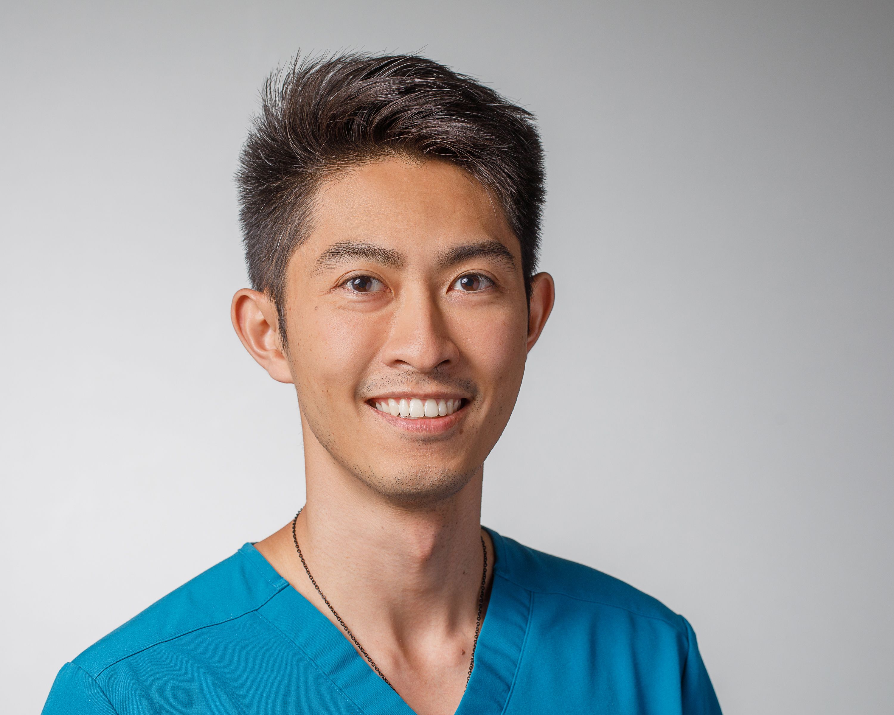
“Each guideline typically requires 5 to 15 systematic reviews, making the process time-consuming (averaging more than 60 weeks) and costly (more than $140,000),” the investigators wrote in Gastroenterology . “One of the most critical yet time-consuming steps in systematic reviews is title and abstract screening. LLMs have the potential to make this step more efficient.”
To test this approach, the investigators developed, validated, and applied a dual-model LLM screening pipeline with human-in-the-loop oversight, focusing on randomized controlled trials in AGA guidelines.
The system was built using the 2021 guideline on moderate-to-severe Crohn’s disease, targeting biologic therapies for induction and maintenance of remission.
Using chain-of-thought prompting and structured inclusion criteria based on the PICO framework, the investigators deployed GPT-4o (OpenAI) and Gemini-1.5-Pro (Google DeepMind) as independent screeners, each assessing titles and abstracts according to standardized logic encoded in JavaScript Object Notation. This approach mimicked a traditional double-reviewer system.
After initial testing, the pipeline was validated in a 2025 update of the same guideline, this time spanning 6 focused clinical questions on advanced therapies and immunomodulators. Results were compared against manual screening by 2 experienced human reviewers, with total screening time documented.
The system was then tested across 4 additional guideline topics: fecal microbiota transplantation (FMT) for irritable bowel syndrome and Clostridioides difficile, gastroparesis, and hepatocellular carcinoma. A final test applied the system to a forthcoming guideline on complications of acute pancreatitis.
Across all topics, the dual-LLM system achieved 100% sensitivity in identifying randomized controlled trials (RCTs). For the 2025 update of the AGA guideline on Crohn’s disease, the models flagged 418 of 4,377 abstracts for inclusion, captur-ing all 25 relevant RCTs in just 48 minutes. Manual screening of the same dataset previously took almost 13 hours.
Comparable accuracy and time savings were observed for the other topics.
The pipeline correctly flagged all 13 RCTs in 4,820 studies on FMT for irritable bowel syndrome, and all 16 RCTs in 5,587 studies on FMT for Clostridioides difficile, requiring 27 and 66 minutes, respectively. Similarly, the system captured all 11 RCTs in 3,919 hepatocellular carcinoma abstracts and all 18 RCTs in 1,578 studies on gastroparesis, completing each task in under 65 minutes. Early testing on the upcoming guideline for pancreatitis yielded similar results.
Cost analysis underscored the efficiency of this approach. At an estimated $175–200 per hour for expert screeners, traditional abstract screening would cost around $2,500 per review, versus approximately $100 for the LLM approach—a 96% reduction.
The investigators cautioned that human oversight remains necessary to verify the relevance of studies flagged by the models. While the system’s sensitivity was consistent, it also selected articles that were ultimately excluded by expert reviewers. Broader validation will be required to assess performance across non-RCT study designs, such as observational or case-control studies, they added.
“As medical literature continues to expand, the integration of artificial intelligence into evidence synthesis processes will become increasingly vital,” Dr. Chung and colleagues wrote. “With further refinement and broader validation, this LLM-based pipeline has the potential to revolutionize evidence synthesis and set a new standard for guideline development.”
This study was funded by National Institutes of Health, National Institute of Diabetes and Digestive and Kidney Diseases. The investigators reported no conflicts of interest.
Ethan Goh, MD, executive director of the Stanford AI Research and Science Evaluation (ARISE) Network, described the AGA pilot as both timely and promising.
“I’m certainly bullish about the use case,” he said in an interview. “Their study design and application is also robust, so I would congratulate them.”

Goh, a general editor for BMJ Digital Health & AI, predicted “huge potential” in the strategy for both clinicians and the general population, who benefit from the most up-to-date guidelines possible.
“I believe that using AI can represent a much faster, more cost effective, efficient way of gathering all these information sources,” he said.
Still, humans will need to be involved in the process.
“[This AI-driven approach] will always need some degree of expert oversight and judgement,” Goh said.
Speaking more broadly about automating study aggregation, Goh said AI may still struggle to determine which studies are most clinically relevant.
“When we use [AI models] to pull out medical references, anecdotally, I don’t think they’re always getting the best ones all the time, or even necessarily the right ones,” he said.
And as AI models grow more impressive, these shortcomings become less apparent, potentially lulling humans into overconfidence.
“Humans are humans,” Goh said. “We get lazy over time. That will be one of the challenges. As the systems get increasingly good, humans start to defer more and more of their judgment to them and say, ‘All right, AI, you’re doing good. Just do 100% automation.’ And then [people] start fact checking or reviewing even less.”
AI could also undermine automated reviews in another way: AI-generated publications that appear genuine, but aren’t, may creep into the dataset.
Despite these concerns, Goh concluded on an optimistic note.
“I think that there are huge ways to use AI, tools, not to replace, but to augment and support human judgment,” he said.
Ethan Goh, MD, is senior research engineer and executive director of the Stanford AI Research and Science Evaluation (ARISE) Network, at Stanford (Calif.) University. He declared no conflicts of interest.
Ethan Goh, MD, executive director of the Stanford AI Research and Science Evaluation (ARISE) Network, described the AGA pilot as both timely and promising.
“I’m certainly bullish about the use case,” he said in an interview. “Their study design and application is also robust, so I would congratulate them.”
Goh, a general editor for BMJ Digital Health & AI, predicted “huge potential” in the strategy for both clinicians and the general population, who benefit from the most up-to-date guidelines possible.
“I believe that using AI can represent a much faster, more cost effective, efficient way of gathering all these information sources,” he said.
Still, humans will need to be involved in the process.
“[This AI-driven approach] will always need some degree of expert oversight and judgement,” Goh said.
Speaking more broadly about automating study aggregation, Goh said AI may still struggle to determine which studies are most clinically relevant.
“When we use [AI models] to pull out medical references, anecdotally, I don’t think they’re always getting the best ones all the time, or even necessarily the right ones,” he said.
And as AI models grow more impressive, these shortcomings become less apparent, potentially lulling humans into overconfidence.
“Humans are humans,” Goh said. “We get lazy over time. That will be one of the challenges. As the systems get increasingly good, humans start to defer more and more of their judgment to them and say, ‘All right, AI, you’re doing good. Just do 100% automation.’ And then [people] start fact checking or reviewing even less.”
AI could also undermine automated reviews in another way: AI-generated publications that appear genuine, but aren’t, may creep into the dataset.
Despite these concerns, Goh concluded on an optimistic note.
“I think that there are huge ways to use AI, tools, not to replace, but to augment and support human judgment,” he said.
Ethan Goh, MD, is senior research engineer and executive director of the Stanford AI Research and Science Evaluation (ARISE) Network, at Stanford (Calif.) University. He declared no conflicts of interest.
, according to a pilot study from the American Gastroenterological Association (AGA).
Faster, cheaper study screening could allow societies to update clinical recommendations more frequently, improving alignment with the latest evidence, lead author Sunny Chung, MD, of Yale School of Medicine, New Haven, Connecticut, and colleagues, reported.
“Each guideline typically requires 5 to 15 systematic reviews, making the process time-consuming (averaging more than 60 weeks) and costly (more than $140,000),” the investigators wrote in Gastroenterology . “One of the most critical yet time-consuming steps in systematic reviews is title and abstract screening. LLMs have the potential to make this step more efficient.”
To test this approach, the investigators developed, validated, and applied a dual-model LLM screening pipeline with human-in-the-loop oversight, focusing on randomized controlled trials in AGA guidelines.
The system was built using the 2021 guideline on moderate-to-severe Crohn’s disease, targeting biologic therapies for induction and maintenance of remission.
Using chain-of-thought prompting and structured inclusion criteria based on the PICO framework, the investigators deployed GPT-4o (OpenAI) and Gemini-1.5-Pro (Google DeepMind) as independent screeners, each assessing titles and abstracts according to standardized logic encoded in JavaScript Object Notation. This approach mimicked a traditional double-reviewer system.
After initial testing, the pipeline was validated in a 2025 update of the same guideline, this time spanning 6 focused clinical questions on advanced therapies and immunomodulators. Results were compared against manual screening by 2 experienced human reviewers, with total screening time documented.
The system was then tested across 4 additional guideline topics: fecal microbiota transplantation (FMT) for irritable bowel syndrome and Clostridioides difficile, gastroparesis, and hepatocellular carcinoma. A final test applied the system to a forthcoming guideline on complications of acute pancreatitis.
Across all topics, the dual-LLM system achieved 100% sensitivity in identifying randomized controlled trials (RCTs). For the 2025 update of the AGA guideline on Crohn’s disease, the models flagged 418 of 4,377 abstracts for inclusion, captur-ing all 25 relevant RCTs in just 48 minutes. Manual screening of the same dataset previously took almost 13 hours.
Comparable accuracy and time savings were observed for the other topics.
The pipeline correctly flagged all 13 RCTs in 4,820 studies on FMT for irritable bowel syndrome, and all 16 RCTs in 5,587 studies on FMT for Clostridioides difficile, requiring 27 and 66 minutes, respectively. Similarly, the system captured all 11 RCTs in 3,919 hepatocellular carcinoma abstracts and all 18 RCTs in 1,578 studies on gastroparesis, completing each task in under 65 minutes. Early testing on the upcoming guideline for pancreatitis yielded similar results.
Cost analysis underscored the efficiency of this approach. At an estimated $175–200 per hour for expert screeners, traditional abstract screening would cost around $2,500 per review, versus approximately $100 for the LLM approach—a 96% reduction.
The investigators cautioned that human oversight remains necessary to verify the relevance of studies flagged by the models. While the system’s sensitivity was consistent, it also selected articles that were ultimately excluded by expert reviewers. Broader validation will be required to assess performance across non-RCT study designs, such as observational or case-control studies, they added.
“As medical literature continues to expand, the integration of artificial intelligence into evidence synthesis processes will become increasingly vital,” Dr. Chung and colleagues wrote. “With further refinement and broader validation, this LLM-based pipeline has the potential to revolutionize evidence synthesis and set a new standard for guideline development.”
This study was funded by National Institutes of Health, National Institute of Diabetes and Digestive and Kidney Diseases. The investigators reported no conflicts of interest.
FROM GASTROENTEROLOGY