User login
Clinical Psychiatry News is the online destination and multimedia properties of Clinica Psychiatry News, the independent news publication for psychiatrists. Since 1971, Clinical Psychiatry News has been the leading source of news and commentary about clinical developments in psychiatry as well as health care policy and regulations that affect the physician's practice.
Dear Drupal User: You're seeing this because you're logged in to Drupal, and not redirected to MDedge.com/psychiatry.
Depression
adolescent depression
adolescent major depressive disorder
adolescent schizophrenia
adolescent with major depressive disorder
animals
autism
baby
brexpiprazole
child
child bipolar
child depression
child schizophrenia
children with bipolar disorder
children with depression
children with major depressive disorder
compulsive behaviors
cure
elderly bipolar
elderly depression
elderly major depressive disorder
elderly schizophrenia
elderly with dementia
first break
first episode
gambling
gaming
geriatric depression
geriatric major depressive disorder
geriatric schizophrenia
infant
ketamine
kid
major depressive disorder
major depressive disorder in adolescents
major depressive disorder in children
parenting
pediatric
pediatric bipolar
pediatric depression
pediatric major depressive disorder
pediatric schizophrenia
pregnancy
pregnant
rexulti
skin care
suicide
teen
wine
section[contains(@class, 'nav-hidden')]
footer[@id='footer']
div[contains(@class, 'pane-pub-article-cpn')]
div[contains(@class, 'pane-pub-home-cpn')]
div[contains(@class, 'pane-pub-topic-cpn')]
div[contains(@class, 'panel-panel-inner')]
div[contains(@class, 'pane-node-field-article-topics')]
section[contains(@class, 'footer-nav-section-wrapper')]
Help! More Clinicians Are Needed to Manage Care for Children With Autism. How About You?
Almost all primary care providers (PCPs) have taken on diagnosing and managing ADHD. With about 12% of school aged children affected, typical PCPs can expect about 240 children with ADHD under their care. Adopting this primary care function has been helped by having clear diagnostic criteria for the three DMS 5 “presentations” of ADHD, open source tools (e.g. Vanderbilts), expectation of collaboration by educators, American Academy of Pediatrics (AAP) guidelines for diagnosis and management, Society for Developmental–Behavioral Pediatrics guidelines for “complex ADHD,” and access to effective medication treatments PCPs can provide (although less so for behavioral ones), cultural acceptance of individuals with ADHD, and especially reliable payment by insurers.
Screening
But what about PCP management of autism spectrum disorder (ASD), now affecting 2.8%, for an expected 60 children under care for each of us? It is more essential because very early detection and entry into evidence-based intervention has long-term benefits for the child and family that are not as crucial for ADHD. While ADHD symptoms may not impact functioning until age 7 or even 12 years of age, signs of ASD usually emerge earlier (by 18 months) but gradually and about 30% after apparently normal development even to age 2 years.
Screening is crucial, but unfortunately not perfect. Recent AAP surveys show that most PCPs screen for autism at the recommended 18 and 24 months. But what happens after that? How many offices are tracking referrals for positive screens for needed evaluations and early intervention? Our data shows that tracking is rarely done and children do not start to get the benefit of early intervention until 4.5 years of age, on average.
Diagnostic Testing
And screening is the easiest part of addressing ASD. Wait times for diagnostic testing can be agonizing months to years. Multiple programs are training PCPs to perform hands-on 10- to 30-minute secondary screening with considerable success. You can become proficient on tools such as STAT (Screening Tool for Autism in Two-Year-Olds), RITA-T (Rapid Interactive Screening Test for Autism in Toddlers), BISCUIT (Baby and Infant Screen for Children with Autism Traits), SORF (Systematic Observation of Red Flags), ADEC (Autism Detection in Early Childhood) or CARS (Childhood Autism Rating Scale) with a few hours of training. Even secondary assessments done virtually by PCPs such as TELE-ASD-PEDS quite accurately predict a verifiable ASD diagnosis for those referred by concerns. Some problems of the reported accuracy of these secondary screening processes have to do with validation in samples of children for whom parents or clinicians already had concern and generally not including many younger children in whom it is so important to detect. Level of confidence of developmental and behavioral pediatricians of the presence of ASD is highly related to ultimate diagnosis. But success with PCPs’ mastering secondary screening has not yet been reported to convince insurers to approve payment for intervention services such as Applied Behavior Analysis (ABA).
Comorbidity
Co-existing conditions affect the majority of patients with ASD (70%), compared with ADHD, but with a broader range and more debilitating and difficult to manage conditions. More medical co-existing issues such as intellectual disability (25%-75%), seizures (12%-26%), motor incoordination (51%), GI conditions (9%-91%), sleep difficulty (50%-80%), sleep apnea, congenital heart disease, avoidant-restrictive food intake disorder, autoimmune disorders, and genetic syndromes (e.g. Fragile X, tuberous sclerosis, Down, Angelman’s, untreated PKU, neurofibromatosis, Klinefelter syndrome) reflect the range of underpinnings of ASD. The need to detect and manage these co-existing issues, besides assessing hearing and vision, makes our skilled involvement and vigilance in ASD care essential. Referring for help from OTs, PTs, speech pathologists, neurologists, psychologists, and special educators as issues in their domains are prioritized is also our responsibility. We must also help families balance utilizing these resources so as to avoid overwhelm.
Anxiety (50%), ADHD (37%-85%), depression (54%), bipolar (7.3%), suicidal ideation (40% starting < 8 years), and emotion dysregulation, familiar to us from our management of ADHD, may develop but are often less well defined and more intractable in ASD, making use of screening tools essential. Using a system like CHADIS that has online pre-visit and monitoring screens delivered based on algorithms for the numerous co-existing conditions, automated handouts, and functions to make and track referral success can facilitate care for this complex chronic condition. Identifying mental health providers with ASD expertise is more difficult, so more management is on us. While medications for these conditions can be beneficial, we need to learn to use lower doses, slower dose increases, and employ problem-solving of side effects with more parent collaboration than for ADHD as children with ASD often cannot self-report effectively. We need to ask about the common ad hoc use of complementary medications and substances (32%-87%) that may be complicating. Of course, these conditions and the caveats of management require more of our time with the patient and family as well as communication with the many other professionals involved. It is important to set our own and our families’ expectations (and schedules) for much more frequent contact and also to bill appropriately with chronic care (99487,89,90) and collaborative care CPT codes (99492,3,4 or G2214).
Behavioral Manifestations
During our care, the often extreme behavioral manifestations of ASD may be the most pressing issues. We need new understanding and skills to sort out and counsel on inflexible, explosive, and sensory triggered behaviors. Just as for ADHD, using the approach of Functional Behavioral Assessment and plans for home as well as school behavior can be key. More difficult in ASD is looking for physical causes, since the child may not provide clear cues because of communication and sensory differences. Conditions common in children with ASD such as constipation, dental caries, otitis, dietary intolerances, allergies, migraine, sleep deficits, menstrual cramps, or fears and changes from puberty manifesting behaviorally are often tricky to sort out.
While the diagnosis of ASD, as for ADHD, does not require any laboratory testing, looking for possible causes is important information for the family and someday may also lead to genetic or other therapies. We need to know that recommendations include screening for Ferritin, Pb, chromosomal microarray and FMR I testing as well as checking that PKU was normal; MECP 2 is indicated in females and symptomatic males; and PTENS testing for children with head circumference greater than 2.5-3 SD. Metabolic and mitochondrial assays are indicated only when symptoms suggest. We need to develop confidence to reserve MRIs or EEGs for cases with abnormal neuro. exams, regression, or history of seizures. It is demanding to keep up with AAP recommendations in this very active area of research.
Interventions
The interventions for ADHD are generally school accommodations and therapies for comorbidities. In contrast, since core social communication skills are the main deficit in ASD, all children screened positive for ASD should be referred for early intervention while awaiting, as well as after, diagnosis. While all states have no or low-cost early intervention, quality and quantity (of hours offered) varies. We should also recommend and try to determine if evidence-based intervention is being provided, such as pivotal response training, UCLA discrete trial therapy, Carbone’s verbal behavior, applied behavior analysis (ABA), Early Start Denver Model, and sometimes music and social skills trainings (effect size 0.42-0.76). Such professional interventions have best evidence with more than 25 hours/week but 15 hours has benefit for higher functioning children. CBT can help anxiety even in younger children. One way for families to provide more hours and more generalizable intervention is coaching by the PLAY Project or DIRFloortime, parent mediated interventions with evidence, some with training both in person or online. Alternative communication training and other condition specific assistance are often needed (e.g. Picture Exchange Communication System for nonverbal children).
While we should already be familiar with writing 504 plan and IEP requests to schools, which also apply to children with ASD, in addition we need to be ready to advise about other legal rights including autism waivers, wraparound services, guardianship, and trust accounts. We can share quality educational materials available online (e.g. from Autism Speaks, SPARK, and Autism Navigator). Social media groups may be supportive, but also may contain disinformation we need to dispel.
Unfortunately, templates, questionnaires, and lack of interdisciplinary referral and communication functions of EHRs don’t support the complexities of care for ASD. While the AAP has guidelines for diagnosis and management and an online toolkit, consider adding a system with an autism-specific module like CHADIS and joining the Autism Care Network or ECHO Autism sessions to get both information and support to take on the evolving critical role of autism care.
Dr. Howard is assistant professor of pediatrics at Johns Hopkins University, Baltimore, and creator of CHADIS. She had no other relevant disclosures. Dr. Howard’s contribution to this publication was as a paid expert to MDedge News. E-mail her at pdnews@mdedge.com.
Better Sleep Tied to Less Loneliness
HOUSTON — Sleep may have a role in driving down rates of loneliness, especially among younger adults.
A study of nearly 2300 participants showed that better sleep health is associated with significantly lower levels of loneliness across ages and that the association is particularly strong in younger individuals.
The US Surgeon General has identified loneliness as “a major public health concern, linked to high rates of negative physical and mental health outcomes,” lead researcher Joseph Dzierzewski, PhD, vice president for research and scientific affairs at the National Sleep Foundation, told this news organization.
“Loneliness is an urgent public health crisis, and there is a pressing need for providers to better understand and treat it,” Dr. Dzierzewski said in a statement.
“Better sleep health might be connected to lower feelings of loneliness by empowering people to engage in social activities, reducing feelings of negative emotions and increasing the likelihood that people interpret interactions in a positive way,” he added.
The findings were presented at SLEEP 2024: 38th Annual Meeting of the Associated Professional Sleep Societies and recently published in an online supplement of the journal Sleep.
Rested, Connected
An American Psychiatric Association poll conducted earlier this year showed 30% of US adults reported feelings of loneliness at least once a week over the past year, and 10% reported feeling lonely every day.
Younger people are more likely to report feeling lonely, with 30% of Americans, aged 18-34 years, feeling lonely every day or several times a week.
While there is growing research identifying a relationship between loneliness and poor sleep in different age groups, few studies have explored ties between social and emotional loneliness and sleep health across the adult lifespan.
In the current study led by Dr. Dzierzewski, 2297 adults (mean age, 44 years; 51% male) completed a validated sleep health questionnaire and loneliness scale.
Linear regression analyses were used to examine the direct associations between sleep health, age, and loneliness. Moderation analyses tested whether the link between sleep health and loneliness differed by age.
On average, the total sleep score was 7.7 (range, 0-12), with higher scores indicating better multidimensional sleep health, and total loneliness scale score was 8.9 (out of 11), indicating moderate levels of loneliness.
Better sleep health and younger age were associated with significantly lower loneliness total scores and social and emotional loneliness subscale scores (all P < .001).
Age significantly moderated the association between sleep health and total (P < .001) and emotional loneliness scores (P < .001) but did not moderate the association between sleep health and social loneliness (P = .034). Better sleep health was associated with lower loneliness across ages, and this association was stronger at younger ages.
“Why younger adults might experience more sleep-related benefits to loneliness than older adults is unknown and intriguing — certainly worth further investigation,” Dr. Dzierzewski said in a conference statement.
Untapped Avenue
Promoting sleep health may be an “untapped avenue” to support efforts and programs that aim to reduce loneliness and increase engagement in all age groups but especially in younger ages, the researchers noted.
Future research should consider monitoring sleep health in programs or interventions that address loneliness, they added.
“Healthcare providers should be aware of the important link between sleep health and loneliness as both sleep and social connections are essential to health and well-being. When sitting across from patients, asking about both sleep health and loneliness might yield important insights into avenues for health promotion,” said Dr. Dzierzewski.
Michael Breus, PhD, clinical psychologist and founder of SleepDoctor.com, who wasn’t involved in the study, is not surprised by the results.
It makes sense that better sleep would lead to less feelings of loneliness, he told this news organization.
Research has shown that when someone is not sleeping well, they “give others a sense of unhappiness, which socially deflects new encounters or even encounters with friends. So social awareness and social initiation would appear to both be affected by sleep quality, therefore potentially leading, at least in part, to loneliness,” he said.
Support for the study was provided by the National Institute on Aging. Dr. Dzierzewski and Dr. Breus had no relevant disclosures.
A version of this article first appeared on Medscape.com.
FROM SLEEP 2024
Emergency Department Visits for Suicide Attempts Rise Across the United States
TOPLINE:
Emergency department (ED) visits in the United States for suicide attempts and intentional self-harm show an increasing trend from 2011 to 2020, with visits being most common among adolescents and the largest increase in visits being seen in adults aged 65 years or older.
METHODOLOGY:
- This study used data from the National Hospital Ambulatory Medical Care Survey, an annual nationwide cross-sectional survey, to track trends in ED visits for suicide attempts and intentional self-harm in the United States from 2011 to 2020.
- Researchers identified visits for suicide attempts and intentional self-harm, along with diagnoses of any co-occurring mental health conditions, using discharge diagnosis codes or reason-for-visit codes.
- The focus was to identify the percentages of ED visits for suicide attempts and intentional self-harm, with analyses done per 100,000 persons and for changes possibly linked to the COVID-19 pandemic in 2019-2020.
TAKEAWAY:
- The number of ED visits owing to suicide attempts and intentional self-harm increased from 1.43 million in 2011-2012 to 5.37 million in 2019-2020 (average annual percent change, 19.5%; 95% confidence interval, 16.9-22.2).
- The rate of ED visits for suicide attempts and intentional self-harm was higher among adolescents and young adults, particularly women, and lower among children.
- Despite a surge in ED visits for self-harm, less than 16% included a mental health evaluation, with visits among patients with mood disorders decreasing by 5.5% annually and those among patients with drug-related disorders increasing by 6.8% annually.
- In 2019-2020, those aged 15-20 years had the highest rate of ED visits (1552 visits per 100,000 persons), with a significant increase seen across all age groups; the largest increase was among those aged 65 years or older.
IN PRACTICE:
“Given that suicide attempts are the single greatest risk factor for suicide, evidence-based management of individuals presenting to emergency departments with suicide attempts and intentional self-harm is a critical component of comprehensive suicide prevention strategies,” the authors wrote.
SOURCE:
The investigation, led by Tanner J. Bommersbach, MD, MPH, Department of Psychiatry and Psychology, Mayo Clinic, Rochester, Minnesota, was published online in The American Journal of Psychiatry.
LIMITATIONS:
Visits for suicide attempts and intentional self-harm were identified based on discharge diagnostic and reason-for-visit codes, which may have led to an underestimation of visits for suicide attempts. ED visits for suicidal vs nonsuicidal self-injury could not be distinguished due to reliance on discharge diagnostic codes. Visits for suicidal ideation, which was not the focus of the study, may have been miscoded as suicide attempts and intentional self-harm.
DISCLOSURES:
No funding source was reported for the study. Some authors received funding grants from various institutions, and one author disclosed receiving honoraria for service as a review committee member and serving as a stakeholder/consultant and as an advisory committee member for some institutes and agencies.
A version of this article appeared on Medscape.com.
DEA Training Mandate: 8 Hours of My Life I’d Like Back
It’s time to renew two of my three narcotic prescribing licenses. For the first time in my career, I’ve waffled on whether the financial outlay to the US Drug Enforcement Agency (DEA) is worth it.
At $888 each, I’ve considered letting two licenses lapse because I only work part-time in Montana. But several friends advised me to keep a “spare” in case I transfer to a new location.
I thought about just paying the fees until I could do a little more research, but there is no mechanism for a refund unless I die within the first year of the 3-year cycle, provide incorrect credit card digits, or accidentally duplicate payments.
The renewal fee is just part of the issue.
Mandatory 8-Hour Training
I also received an alert about the requirement for more “narcotics prescribing education” thanks to the Medication Access and Training Expansion Act (MATE).
The requirement seems counterintuitive because opioid prescribing has decreased for the 10th consecutive year, according to the AMA Overdose Epidemic Report. The continuing rise in overdose deaths is largely due to illegitimate manufacturing of synthetic opioids.
I’ve written zero outpatient narcotics prescriptions in the past 6 years, and I’ve written very few in my 33 years of practice. My use is limited to intravenous morphine for flash pulmonary edema or refractory angina, but unless you graduated from a training program within 5 years of the June 2023 mandate or are boarded in addiction medicine, there is no way to escape the 8-hour education requirement.
The problem is that these courses are never just 8 hours in duration. After signing up for one such CME course that cost $150, I was still dying of boredom and at risk for DVT 4 days later. That’s how long it took to sit through.
Instead of the 30 seconds it should have taken to review the simple instructions to deliver Narcan, there were scores of screens followed by juvenile quizlets and cartoons. All but about 2 hours out of the 4 days is now relegated to that category of “hours of my life that I can never get back.” Additionally, none of that mandatory “education” will change my prescribing habits one whit.
And beware the penalty. 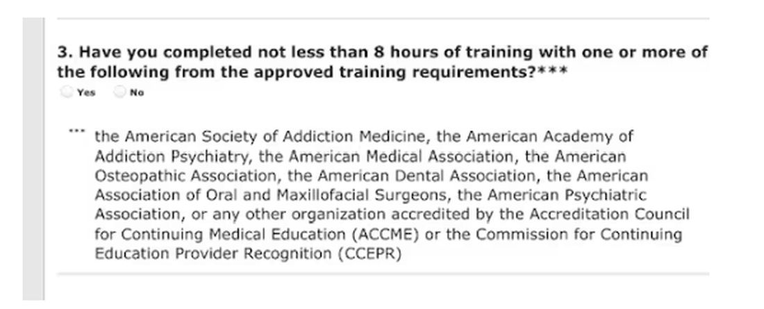
Of course, I would always be truthful when asked to check the box on the DEA renewal application attesting to my having completed the required education. On the outside chance that you plan to check the yes box without completing the relevant courses, those found guilty of such false claims could be fined up to $250,000 and subject to “not more than four years in prison,” or both. Yikes! 
Larry Houck, a former DEA investigator, explained that “[t]here are lot of people who are coming up for renewal and log on but still don’t know this is a requirement.” Neither ignorance nor complacency is an acceptable defense.
Changes Needed
The only good thing that came of those 4 long days of opioid education was a motivation to drive change in our current licensing and educational experience. Why not use this opportunity to reform the DEA-physician/prescriber relationship?
The educational requirements should be curtailed for those of us who do not provide outpatient narcotic prescriptions even if we use inpatient opioids. Meds with low abuse potential should be rescheduled to minimize who gets caught in the broad net of the education requirement.
We should reduce overregulation of the legitimate prescribers by lowering, instead of increasing, licensing fees. We should change to a single license number that covers every state. In this digital age, there is no legitimate excuse to prevent this from happening.
After all, the settlements from opioid manufacturers and distributors will in time total $50 billion. It seems that at least some of the responsibilities of the DEA could shift to states, cities, and towns.
My friend Siamak Karimian, MD, who provides locum services in multiple states, pays for seven active DEA licenses every 3 years. He pointed out the hypocrisy in the current regulatory system: “It’s funny that you can have only one DEA or state license and work for the government in all other states or territories with no limits, including the VA, Indian healthcare systems, or prison systems.”
All other prescribers require a separate DEA number for every state. Ultimately, you’d think tracking prescriptions for a single DEA number should be far simpler than tracking someone with seven.
Competent physicians not guilty of criminal overprescribing seem to be the last to be considered in nearly every healthcare endeavor these days. It would be refreshing if they would reduce our fees and prevent this waste of our time.
And while we are at it, perhaps a more fitting punishment is due for Richard Sackler and all the Purdue Pharma–affiliated family members. The Sacklers will pay out $6 billion in exchange for immunity against civil litigation. That doesn’t seem like much when they are worth $11 billion.
Perhaps they should be made to take an 8-hour course on opioid prescribing, annually and in perpetuity. Let’s see them complete a few quizlets and sit through screens of instruction on how to administer Naloxone. Of course, that would be a mild punishment for those who manufactured a drug that killed hundreds of thousands. But it would be a start.
Dr. Walton-Shirley, a clinical cardiologist in Nashville, Tennessee, has disclosed no relevant financial relationships.
A version of this article appeared on Medscape.com.
It’s time to renew two of my three narcotic prescribing licenses. For the first time in my career, I’ve waffled on whether the financial outlay to the US Drug Enforcement Agency (DEA) is worth it.
At $888 each, I’ve considered letting two licenses lapse because I only work part-time in Montana. But several friends advised me to keep a “spare” in case I transfer to a new location.
I thought about just paying the fees until I could do a little more research, but there is no mechanism for a refund unless I die within the first year of the 3-year cycle, provide incorrect credit card digits, or accidentally duplicate payments.
The renewal fee is just part of the issue.
Mandatory 8-Hour Training
I also received an alert about the requirement for more “narcotics prescribing education” thanks to the Medication Access and Training Expansion Act (MATE).
The requirement seems counterintuitive because opioid prescribing has decreased for the 10th consecutive year, according to the AMA Overdose Epidemic Report. The continuing rise in overdose deaths is largely due to illegitimate manufacturing of synthetic opioids.
I’ve written zero outpatient narcotics prescriptions in the past 6 years, and I’ve written very few in my 33 years of practice. My use is limited to intravenous morphine for flash pulmonary edema or refractory angina, but unless you graduated from a training program within 5 years of the June 2023 mandate or are boarded in addiction medicine, there is no way to escape the 8-hour education requirement.
The problem is that these courses are never just 8 hours in duration. After signing up for one such CME course that cost $150, I was still dying of boredom and at risk for DVT 4 days later. That’s how long it took to sit through.
Instead of the 30 seconds it should have taken to review the simple instructions to deliver Narcan, there were scores of screens followed by juvenile quizlets and cartoons. All but about 2 hours out of the 4 days is now relegated to that category of “hours of my life that I can never get back.” Additionally, none of that mandatory “education” will change my prescribing habits one whit.
And beware the penalty.
Of course, I would always be truthful when asked to check the box on the DEA renewal application attesting to my having completed the required education. On the outside chance that you plan to check the yes box without completing the relevant courses, those found guilty of such false claims could be fined up to $250,000 and subject to “not more than four years in prison,” or both. Yikes!
Larry Houck, a former DEA investigator, explained that “[t]here are lot of people who are coming up for renewal and log on but still don’t know this is a requirement.” Neither ignorance nor complacency is an acceptable defense.
Changes Needed
The only good thing that came of those 4 long days of opioid education was a motivation to drive change in our current licensing and educational experience. Why not use this opportunity to reform the DEA-physician/prescriber relationship?
The educational requirements should be curtailed for those of us who do not provide outpatient narcotic prescriptions even if we use inpatient opioids. Meds with low abuse potential should be rescheduled to minimize who gets caught in the broad net of the education requirement.
We should reduce overregulation of the legitimate prescribers by lowering, instead of increasing, licensing fees. We should change to a single license number that covers every state. In this digital age, there is no legitimate excuse to prevent this from happening.
After all, the settlements from opioid manufacturers and distributors will in time total $50 billion. It seems that at least some of the responsibilities of the DEA could shift to states, cities, and towns.
My friend Siamak Karimian, MD, who provides locum services in multiple states, pays for seven active DEA licenses every 3 years. He pointed out the hypocrisy in the current regulatory system: “It’s funny that you can have only one DEA or state license and work for the government in all other states or territories with no limits, including the VA, Indian healthcare systems, or prison systems.”
All other prescribers require a separate DEA number for every state. Ultimately, you’d think tracking prescriptions for a single DEA number should be far simpler than tracking someone with seven.
Competent physicians not guilty of criminal overprescribing seem to be the last to be considered in nearly every healthcare endeavor these days. It would be refreshing if they would reduce our fees and prevent this waste of our time.
And while we are at it, perhaps a more fitting punishment is due for Richard Sackler and all the Purdue Pharma–affiliated family members. The Sacklers will pay out $6 billion in exchange for immunity against civil litigation. That doesn’t seem like much when they are worth $11 billion.
Perhaps they should be made to take an 8-hour course on opioid prescribing, annually and in perpetuity. Let’s see them complete a few quizlets and sit through screens of instruction on how to administer Naloxone. Of course, that would be a mild punishment for those who manufactured a drug that killed hundreds of thousands. But it would be a start.
Dr. Walton-Shirley, a clinical cardiologist in Nashville, Tennessee, has disclosed no relevant financial relationships.
A version of this article appeared on Medscape.com.
‘Shockingly High’ Rate of TBI in Older Adults
TOPLINE:
, a new study showed.
METHODOLOGY:
- Researchers analyzed data from approximately 9200 Medicare enrollees who were part of the Health and Retirement Study (HRS), aged 65 years and older, from 2000 to 2018.
- The baseline date was the date of the first age eligible HRS core interview in the community in 2000 or later.
- Incident TBI cases came from an updated list of the International Classification of Diseases (ICD), 9th and 10th edition codes, from the Defense and Veterans Brain Injury Center and the Armed Forces Health Surveillance Branch for TBI surveillance.
- Codes corresponded with emergency department, CT, and/or fMRI visits.
TAKEAWAY:
- Almost 13% of older individuals (n = 797) experienced TBI during the study, highlighting its significant prevalence in this population.
- Older adults (mean age at baseline, 75 years) who experienced TBI during the study period were more likely to be women and White individuals as well as individuals having higher levels of education and normal cognition (P < .001), challenging previous assumptions about risk factors.
- The study underscored the need for targeted interventions and research focused on TBI prevention and postdischarge care in older adults.
IN PRACTICE:
“The number of people 65 and older with TBI is shockingly high,” senior author Raquel Gardner, MD, said in a press release. “We need evidence-based guidelines to inform postdischarge care of this very large Medicare population and more research on post-TBI dementia prevention and repeat injury prevention.”
SOURCE:
The study was led by Erica Kornblith, PhD, of the University of California, San Francisco. It was published online in JAMA Network Open.
LIMITATIONS:
The study’s reliance on ICD codes for TBI identification may not capture the full spectrum of TBI severity. Self-reported data on sociodemographic factors may have introduced bias, affecting the accuracy of associations with TBI incidence. In addition, the findings’ generalizability may be limited due to the study’s focus on Medicare enrollees, potentially excluding those from diverse socioeconomic backgrounds.
DISCLOSURES:
The study was funded by the Alzheimer’s Association, the US Department of Veterans Affairs, the National Institute on Aging, and the Department of Defense. Disclosures are noted in the original study.
This article was created using several editorial tools, including AI, as part of the process. Human editors reviewed this content before publication.
A version of this article appeared on Medscape.com.
Sharp Rise in US Pediatric ADHD Diagnoses
TOPLINE:
METHODOLOGY:
- Researchers used 2022 data from the National Survey of Children’s Health to estimate the prevalence of ever-diagnosed and current ADHD among US children between the ages of 3 and 18 years.
- They also estimated, among children with current ADHD, the severity of the condition and the presence of current co-occurring disorders and the receipt of medication and behavioral treatments.
- The researchers calculated overall weighted estimates as well as estimates for specific demographic and clinical subgroups (n = 45,169).
TAKEAWAY:
- The number of children who had ever received an ADHD diagnosis increased from 6.1 million in 2016 to 7.1 million in 2022, and the number with current ADHD increased from 5.4 million to 6.5 million.
- Of those with current ADHD in 2022, 58.1% had moderate or severe ADHD, and 77.9% had at least one co-occurring disorder.
- A total of 53.6% had received ADHD medication, 44.4% had received behavioral treatment in the past year, and 30.1% had received no ADHD-specific treatment.
- A similar percentage of children with ADHD were receiving behavioral treatment in 2022 as in 2016 (44.4% vs 46.7%, respectively), but treatment with ADHD medication was lower in 2022 than in 2016 (53.6% vs 62.0%, respectively).
IN PRACTICE:
The estimates “can be used by clinicians to understand current ADHD diagnosis and treatment utilization patterns to inform clinical practice, such as accounting for the frequency and management of co-occurring conditions and considering the notable percentage of children with ADHD not currently receiving ADHD treatment,” and can be used by policymakers, practitioners, and others “to plan for the needs of children with ADHD, such as by ensuring access to care and services for ADHD,” investigators wrote.
SOURCE:
Melissa L. Danielson, of the National Center on Birth Defects and Developmental Disabilities, Centers for Disease Control and Prevention, led the study, which was published online in the Journal of Clinical Child & Adolescent Psychology.
LIMITATIONS:
Indicators reported in the analysis were on the basis of the parent report, which may be limited by recall and reporting decisions and were not validated against medical records or clinical judgment. Moreover, details about the types of treatment were not included.
DISCLOSURES:
The work was authorized as part of the contributor’s official duties as an employee of the US Government, and therefore is a work of the US Government. The authors declared no relevant financial relationships.
A version of this article appeared on Medscape.com.
Novel Method Able to Predict if, When, Dementia Will Develop
Novel, noninvasive testing is able to predict dementia onset with 80% accuracy up to 9 years before clinical diagnosis.
The results suggest resting-state functional MRI (rs-fMRI) could be used to identify a neural network signature of dementia risk early in the pathological course of the disease, an important advance as disease-modifying drugs such as those targeting amyloid beta are now becoming available.
“The brain has been changing for a long time before people get symptoms of dementia, and if we’re very precise about how we do it, we can actually, in principle, detect those changes, which could be really exciting,” study investigator Charles R. Marshall, PhD, professor of clinical neurology, Centre for Preventive Neurology, Wolfson Institute of Population Health, Queen Mary University of London, London, England, told this news organization.
“This could become a platform for screening people for risk status in the future, and it could one day make all the difference in terms of being able to prevent dementia,” he added.
The findings were published online in Nature Mental Health.
The rs-fMRI measures fluctuations in blood oxygen level–dependent signals across the brain, which reflect functional connectivity.
Brain regions commonly implicated in altered functional connectivity in Alzheimer’s disease (AD) are within the default-mode network (DMN). This is the group of regions “connecting with each other and communicating with each other when someone is just lying in an MRI scanner doing nothing, which is how it came to be called the default-mode network,” explained Dr. Marshall.
The DMN encompasses the medial prefrontal cortex, posterior cingulate cortex or precuneus, and bilateral inferior parietal cortices, as well as supplementary brain regions including the medial temporal lobes and temporal poles.
This network is believed to be selectively vulnerable to AD neuropathology. “Something about that network starts to be disrupted in the very earliest stages of Alzheimer’s disease,” said Dr. Marshall.
While this has been known for some time, “what we’ve not been able to do before is build a precise enough model of how the network is connected to be able to tell whether individual participants were going to get dementia or not,” he added.
The investigators used data from the UK Biobank, a large-scale biomedical database and research resource containing genetic and health information from about a half a million UK volunteer participants.
The analysis included 103 individuals with dementia (22 with prevalent dementia and 81 later diagnosed with dementia over a median of 3.7 years) and 1030 matched participants without dementia. All participants had MRI imaging between 2006 and 2010.
The total sample had a mean age of 70.4 years at the time of MRI data acquisition. For each participant, researchers extracted relevant data from 10 predefined regions of interest in the brain, which together defined their DMN. This included two midline regions and four regions in each hemisphere.
Greater Predictive Power
Researchers built a model using an approach related to how brain regions communicate with each other. “The model sort of incorporates what we know about how the changes that you see on a functional MRI scan relate to changes in the firing of brain cells, in a very precise way,” said Dr. Marshall.
The researchers then used a machine learning approach to develop a model for effective connectivity, which describes the causal influence of one brain region over another. “We trained a machine learning tool to recognize what a dementia-like pattern of connectivity looks like,” said Dr. Marshall.
Investigators controlled for potential confounders, including age, sex, handedness, in-scanner head motion, and geographical location of data acquisition.
The model was able to determine the difference in brain connectivity patterns between those who would go on to develop dementia and those who would not, with an accuracy of 82% up to 9 years before an official diagnosis was made.
When the researchers trained a model to use brain connections to predict time to diagnosis, the predicted time to diagnosis and actual time to diagnosis were within about 2 years.
This effective connectivity approach has much more predictive power than memory test scores or brain structural measures, said Dr. Marshall. “We looked at brain volumes and they performed very poorly, only just better than tossing a coin, and the same with cognitive test scores, which were only just better than chance.”
As for markers of amyloid beta and tau in the brain, these are “very useful diagnostically” but only when someone has symptoms, said Dr. Marshall. He noted people live for years with these proteins without developing dementia symptoms.
“We wouldn’t necessarily want to expose somebody who has a brain full of amyloid but was not going to get symptoms for the next 20 years to a treatment, but if we knew that person was highly likely to develop symptoms of dementia in the next 5 years, then we probably would,” he said.
Dr. Marshall believes the predictive power of all these diagnostic tools could be boosted if they were used together.
Potential for Early Detection, Treatment
Researchers examined a number of modifiable dementia risk factors, including hearing loss, depression, hypertension, and physical inactivity. They found self-reported social isolation was the only variable that showed a significant association with effective connectivity, meaning those who are socially isolated were more likely to have a “dementia-like” pattern of DMN effective connectivity. This finding suggests social isolation is a cause, rather than a consequence, of dementia.
The study also revealed associations between DMN effective connectivity and AD polygenic risk score, derived from meta-analysis of multiple external genome-wide association study sources.
A predictive tool that uses rs-fMRI could also help select participants at a high risk for dementia to investigate potential treatments. “There’s good reason to think that if we could go in earlier with, for example, anti-amyloid treatments, they’re more likely to be effective,” said Dr. Marshall.
The new test might eventually have value as a population screening tool, something akin to colon cancer screening, he added. “We don’t send everyone for a colonoscopy; you do a kind of pre-screening test at home, and if that’s positive, then you get called in for a colonoscopy.”
The researchers looked at all-cause dementia and not just AD because dementia subtype diagnoses in the UK Biobank “are not at all reliable,” said Dr. Marshall.
Study limitations included the fact that UK Biobank participants are healthier and less socioeconomically deprived than the general population and are predominantly White. Another study limitation was that labeling of cases and controls depended on clinician coding rather than on standardized diagnostic criteria.
Kudos, Caveats
In a release from the Science Media Center, a nonprofit organization promoting voices and views of the scientific community, Sebastian Walsh, National Institute for Health and Care Research doctoral fellow in Public Health Medicine, University of Cambridge, Cambridge, England, said the results are “potentially exciting,” and he praised the way the team conducted the study.
However, he noted some caveats, including the small sample size, with only about 100 people with dementia, and the relatively short time between the brain scan and diagnosis (an average of 3.7 years).
Dr. Walsh emphasized the importance of replicating the findings “in bigger samples with a much longer delay between scan and onset of cognitive symptoms.”
He also noted the average age of study participants was 70 years, whereas the average age at which individuals in the United Kingdom develop dementia is mid to late 80s, “so we need to see these results repeated for more diverse and older samples.”
He also noted that MRI scans are expensive, and the approach used in the study needs “a high-quality scan which requires people to keep their head still.”
Also commenting, Andrew Doig, PhD, professor, Division of Neuroscience, the University of Manchester, Manchester, England, said the MRI connectivity method used in the study might form part of a broader diagnostic approach.
“Dementia is a complex condition, and it is unlikely that we will ever find one simple test that can accurately diagnose it,” Dr. Doig noted. “Within a few years, however, there is good reason to believe that we will be routinely testing for dementia in middle-aged people, using a combination of methods, such as a blood test, followed by imaging.”
“The MRI connectivity method described here could form part of this diagnostic platform. We will then have an excellent understanding of which people are likely to benefit most from the new generation of dementia drugs,” he said.
Dr. Marshall and Dr. Walsh reported no relevant disclosures. Dr. Doig reported that he is a founder, shareholder, and consultant for PharmaKure Ltd, which is developing new diagnostics for neurodegenerative diseases using blood biomarkers.
A version of this article first appeared on Medscape.com.
Chronotherapy: Why Timing Drugs to Our Body Clocks May Work
Do drugs work better if taken by the clock?
A new analysis published in The Lancet journal’s eClinicalMedicine suggests: Yes, they do — if you consider the patient’s individual body clock. The study is the first to find that timing blood pressure drugs to a person’s personal “chronotype” — that is, whether they are a night owl or an early bird — may reduce the risk for a heart attack.
The findings represent a significant advance in the field of circadian medicine or “chronotherapy” — timing drug administration to circadian rhythms. A growing stack of research suggests this approach could reduce side effects and improve the effectiveness of a wide range of therapies, including vaccines, cancer treatments, and drugs for depression, glaucoma, pain, seizures, and other conditions. Still, despite decades of research, time of day is rarely considered in writing prescriptions.
“We are really just at the beginning of an exciting new way of looking at patient care,” said Kenneth A. Dyar, PhD, whose lab at Helmholtz Zentrum München’s Institute for Diabetes and Cancer focuses on metabolic physiology. Dr. Dyar is co-lead author of the new blood pressure analysis.
“Chronotherapy is a rapidly growing field,” he said, “and I suspect we are soon going to see more and more studies focused on ‘personalized chronotherapy,’ not only in hypertension but also potentially in other clinical areas.”
The ‘Missing Piece’ in Chronotherapy Research
Blood pressure drugs have long been chronotherapy’s battleground. After all, blood pressure follows a circadian rhythm, peaking in the morning and dropping at night.
That healthy overnight dip can disappear in people with diabetes, kidney disease, and obstructive sleep apnea. Some physicians have suggested a bed-time dose to restore that dip. But studies have had mixed results, so “take at bedtime” has become a less common recommendation in recent years.
But the debate continued. After a large 2019 Spanish study found that bedtime doses had benefits so big that the results drew questions, an even larger, 2022 randomized, controlled trial from the University of Dundee in Dundee, Scotland — called the TIME study — aimed to settle the question.
Researchers assigned over 21,000 people to take morning or night hypertension drugs for several years and found no difference in cardiovascular outcomes.
“We did this study thinking nocturnal blood pressure tablets might be better,” said Thomas MacDonald, MD, professor emeritus of clinical pharmacology and pharmacoepidemiology at the University of Dundee and principal investigator for the TIME study and the recent chronotype analysis. “But there was no difference for heart attacks, strokes, or vascular death.”
So, the researchers then looked at participants’ chronotypes, sorting outcomes based on whether the participants were late-to-bed, late-to-rise “night owls” or early-to-bed, early-to-rise “morning larks.”
Their analysis of these 5358 TIME participants found the following results: Risk for hospitalization for a heart attack was at least 34% lower for “owls” who took their drugs at bedtime. By contrast, owls’ heart attack risk was at least 62% higher with morning doses. For “larks,” the opposite was true. Morning doses were associated with an 11% lower heart attack risk and night doses with an 11% higher risk, according to supplemental data.
The personalized approach could explain why some previous chronotherapy studies have failed to show a benefit. Those studies did not individualize drug timing as this one did. But personalization could be key to circadian medicine’s success.
“Our ‘internal personal time’ appears to be an important variable to consider when dosing antihypertensives,” said co-lead author Filippo Pigazzani, MD, PhD, clinical senior lecturer and honorary consultant cardiologist at the University of Dundee School of Medicine. “Chronotherapy research has been going on for decades. We knew there was something important with time of day. But researchers haven’t considered the internal time of individual people. I think that is the missing piece.”
The analysis has several important limitations, the researchers said. A total of 95% of participants were White. And it was an observational study, not a true randomized comparison. “We started it late in the original TIME study,” Dr. MacDonald said. “You could argue we were reporting on those who survived long enough to get into the analysis.” More research is needed, they concluded.
Looking Beyond Blood Pressure
What about the rest of the body? “Almost all the cells of our body contain ‘circadian clocks’ that are synchronized by daily environmental cues, including light-dark, activity-rest, and feeding-fasting cycles,” said Dr. Dyar.
An estimated 50% of prescription drugs hit targets in the body that have circadian patterns. So, experts suspect that syncing a drug with a person’s body clock might increase effectiveness of many drugs.
A handful of US Food and Drug Administration–approved drugs already have time-of-day recommendations on the label for effectiveness or to limit side effects, including bedtime or evening for the insomnia drug Ambien, the HIV antiviral Atripla, and cholesterol-lowering Zocor. Others are intended to be taken with or after your last meal of the day, such as the long-acting insulin Levemir and the cardiovascular drug Xarelto. A morning recommendation comes with the proton pump inhibitor Nexium and the attention-deficit/hyperactivity disorder drug Ritalin.
Interest is expanding. About one third of the papers published about chronotherapy in the past 25 years have come out in the past 5 years. The May 2024 meeting of the Society for Research on Biological Rhythms featured a day-long session aimed at bringing clinicians up to speed. An organization called the International Association of Circadian Health Clinics is trying to bring circadian medicine findings to clinicians and their patients and to support research.
Moreover, while recent research suggests minding the clock could have benefits for a wide range of treatments, ignoring it could cause problems.
In a Massachusetts Institute of Technology study published in April in Science Advances, researchers looked at engineered livers made from human donor cells and found more than 300 genes that operate on a circadian schedule, many with roles in drug metabolism. They also found that circadian patterns affected the toxicity of acetaminophen and atorvastatin. Identifying the time of day to take these drugs could maximize effectiveness and minimize adverse effects, the researchers said.
Timing and the Immune System
Circadian rhythms are also seen in immune processes. In a 2023 study in The Journal of Clinical Investigation of vaccine data from 1.5 million people in Israel, researchers found that children and older adults who got their second dose of the Pfizer mRNA COVID vaccine earlier in the day were about 36% less likely to be hospitalized with SARS-CoV-2 infection than those who got an evening shot.
“The sweet spot in our data was somewhere around late morning to late afternoon,” said lead researcher Jeffrey Haspel, MD, PhD, associate professor of medicine in the division of pulmonary and critical care medicine at Washington University School of Medicine in St. Louis.
In a multicenter, 2024 analysis of 13 studies of immunotherapy for advanced cancers in 1663 people, researchers found treatment earlier in the day was associated with longer survival time and longer survival without cancer progression.
“Patients with selected metastatic cancers seemed to largely benefit from early [time of day] infusions, which is consistent with circadian mechanisms in immune-cell functions and trafficking,” the researchers noted. But “retrospective randomized trials are needed to establish recommendations for optimal circadian timing.”
Other research suggests or is investigating possible chronotherapy benefits for depression, glaucoma, respiratory diseases, stroke treatment, epilepsy, and sedatives used in surgery. So why aren’t healthcare providers adding time of day to more prescriptions? “What’s missing is more reliable data,” Dr. Dyar said.
Should You Use Chronotherapy Now?
Experts emphasize that more research is needed before doctors use chronotherapy and before medical organizations include it in treatment recommendations. But for some patients, circadian dosing may be worth a try:
Night owls whose blood pressure isn’t well controlled. Dr. Dyar and Dr. Pigazzani said night-time blood pressure drugs may be helpful for people with a “late chronotype.” Of course, patients shouldn’t change their medication schedule on their own, they said. And doctors may want to consider other concerns, like more overnight bathroom visits with evening diuretics.
In their study, the researchers determined participants’ chronotype with a few questions from the Munich Chronotype Questionnaire about what time they fell asleep and woke up on workdays and days off and whether they considered themselves “morning types” or “evening types.” (The questions can be found in supplementary data for the study.)
If a physician thinks matching the timing of a dose with chronotype would help, they can consider it, Dr. Pigazzani said. “However, I must add that this was an observational study, so I would advise healthcare practitioners to wait for our data to be confirmed in new RCTs of personalized chronotherapy of hypertension.”
Children and older adults getting vaccines. Timing COVID shots and possibly other vaccines from late morning to mid-afternoon could have a small benefit for individuals and a bigger public-health benefit, Dr. Haspel said. But the most important thing is getting vaccinated. “If you can only get one in the evening, it’s still worthwhile. Timing may add oomph at a public-health level for more vulnerable groups.”
A version of this article appeared on Medscape.com.
Antidepressants and Dementia Risk: New Data
TOPLINE:
Taking antidepressants in midlife was not associated with an increased risk of subsequent Alzheimer’s disease (AD) or AD-related dementias (ADRD), data from a large prospective study of US veterans show.
METHODOLOGY:
- Investigators analyzed data from 35,200 US veterans aged ≥ 55 years diagnosed with major depressive disorder from January 1, 2000, to June 1, 2022, and followed them for ≤ 20 years to track subsequent AD/ADRD diagnoses.
- Health information was pulled from electronic health records of the Veterans Health Administration (VHA) Corporate Data Warehouse, and veterans had to be at the VHA for ≥ 1 year before diagnosis.
- Participants were considered to be exposed to an antidepressant when a prescription lasted ≥ 3 months.
TAKEAWAY:
- A total of 32,500 individuals were diagnosed with MDD. The mean age was 65 years, and 91% were men. 17,000 patients received antidepressants for a median duration of 4 years. Median follow-up time was 3.2 years.
- There was no significant association between antidepressant exposure and the risk for AD/ADRD (events = 1056; hazard ratio, 0.93; 95% CI, 0.80-1.08) vs no exposure.
- In a subgroup analysis, investigators found no significant link between different classes of antidepressants and dementia risk. These included selective serotonin reuptake inhibitors, norepinephrine and dopamine reuptake inhibitors, and serotonin-norepinephrine reuptake inhibitors.
- Investigators emphasized the need for further research, particularly in populations with a larger representation of female patients.
IN PRACTICE:
“A possibility for the conflicting results in retrospective studies is that the heightened risk identified in participants on antidepressants may be attributed to depression itself, rather than the result of a potential pharmacological action. So, this and other clinical confounding factors need to be taken into account,” the investigators noted.
SOURCE:
The study was led by Jaime Ramos-Cejudo, PhD, VA Boston Healthcare System, Boston. It was published online May 8 in Alzheimer’s & Dementia.
LIMITATIONS:
The cohort’s relatively young age limited the number of dementia cases captured. Data from supplemental insurance, including Medicare, were not included, potentially limiting outcome capture.
DISCLOSURES:
The study was supported by the National Institutes of Health and the National Alzheimer’s Coordinating Center. The authors declared no conflicts of interest.
This article was created using several editorial tools, including AI, as part of the process. Human editors reviewed this content before publication.
A version of this article appeared on Medscape.com.
PTSD Rates Soar Among College Students
TOPLINE:
Posttraumatic stress disorder (PTSD) rates among college students more than doubled between 2017 and 2022, new data showed. Rates of acute stress disorder (ASD) also increased during that time.
METHODOLOGY:
- Researchers conducted five waves of cross-sectional study from 2017 to 2022, involving 392,377 participants across 332 colleges and universities.
- The study utilized the Healthy Minds Study data, ensuring representativeness by applying sample weights based on institutional demographics.
- Outcome variables were diagnoses of PTSD and ASD, confirmed by healthcare practitioners, with statistical analysis assessing change in odds of estimated prevalence during 2017-2022.
TAKEAWAY:
- The prevalence of PTSD among US college students increased from 3.4% in 2017-2018 to 7.5% in 2021-2022.
- ASD diagnoses also rose from 0.2% in 2017-2018 to 0.7% in 2021-2022, with both increases remaining statistically significant after adjusting for demographic differences.
- Investigators noted that these findings underscore the need for targeted, trauma-informed intervention strategies in college settings.
IN PRACTICE:
“These trends highlight the escalating mental health challenges among college students, which is consistent with recent research reporting a surge in psychiatric diagnoses,” the authors wrote. “Factors contributing to this rise may include pandemic-related stressors (eg, loss of loved ones) and the effect of traumatic events (eg, campus shootings and racial trauma),” they added.
SOURCE:
The study was led by Yusen Zhai, PhD, University of Alabama at Birmingham. It was published online on May 30, 2024, in JAMA Network Open.
LIMITATIONS:
The study’s reliance on self-reported data and single questions for diagnosed PTSD and ASD may have limited the accuracy of the findings. The retrospective design and the absence of longitudinal follow-up may have restricted the ability to infer causality from the observed trends.
DISCLOSURES:
No disclosures were reported. No funding information was available.
This article was created using several editorial tools, including AI, as part of the process. Human editors reviewed this content before publication.
A version of this article appeared on Medscape.com.