User login
New York GI Links Health Equity and CRC Screening
Pascale M. White, MD, MBA, MS never tires of excising precancerous polyps.
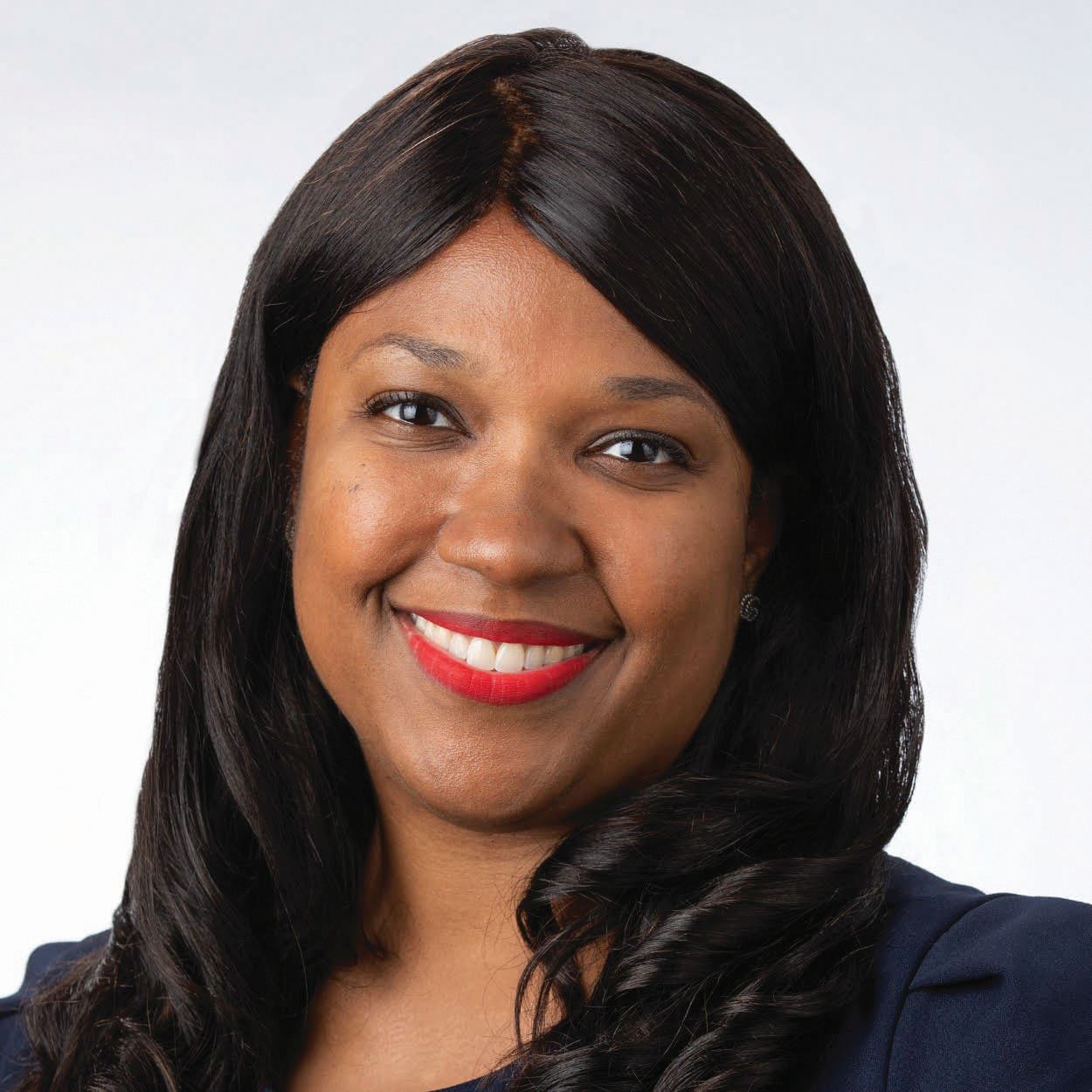
“To know that I have removed something that could have been potentially dangerous to this patient in years to come, that wasn’t causing any symptoms but silently lurking there” is a great feeling, said Dr. White, an associate professor with dual appointments in the divisions of gastroenterology and liver diseases at the Icahn School of Medicine at Mount Sinai, New York.
“When I do procedures, I always go in with the mindset that this could be a lifesaving procedure for this patient. And that definitely keeps me excited about the field,” she said.
Colorectal cancer is preventable, but when it comes to screening, there are large health disparities. African Americans are 20% more likely to get diagnosed with colorectal cancer and 40% more likely to die from the disease. “Knowing that there are low screening rates among this population, there’s a lot of work to be done with mitigating those disparities,” said Dr. White, who has made it her life’s work to expand access to care and address health inequities.
Dr. White is an inaugural director of Health Equity in Action for Liver and Digestive Diseases (HEALD) and an inaugural fellow of the United Hospital Fund’s Health Equity Fellowship. In 2025, she received the AGA-Pfizer Beacon of Hope Award, which celebrates three women in the GI field who have played a key role in advancing gender and health equity in medicine.
“Through the United Hospital Fund’s Health Equity Fellowship, I have partnered with an East Harlem community health center to conduct seminars and tailor a one-page shared decision tool for colorectal cancer screening to jumpstart discussions on screening choices between patients and providers,” said Dr. White.
In an interview, she offered more details about her mission to connect with communities to improve screening rates for colorectal cancer.
Can you discuss your work with HEALD?
Dr. White: HEALD is a growing initiative to identify and address any access barriers to our screening programs. At this time, I’m working to identify how patients are getting referred to us in our division for colorectal cancer screening and how we can create a more streamlined and robust pathway for patients in the community, namely at federally qualified health centers in East Harlem.
You co-founded the Association of Black Gastroenterologists and Hepatologists (ABGH) in 2021. What are you hoping to accomplish with this organization?
Dr. White: ABGH was co-founded by eleven of us from across the country for the purpose of addressing health care disparities in GI and liver diseases that disproportionately affect Black patient populations. Our mission is to promote health equity, advance science and develop the careers of Black gastroenterologists, hepatologists, and scientists.
Our mentorship program is one way we give back to incoming residents who are interested in pursuing a career in GI. The Nurturing, Excelling, and Unifying Sisters in Medicine (NEXUS) conference centers the perspectives of Black women in medicine from all specialties. The ABGH Summit is an educational conference that features renowned experts in the health equity space.
But at the center of it all is our community outreach. When we started the organization after Chadwick Boseman’s death during the height of the COVID pandemic, all our community events were held over Zoom. Now with our in-person events you can feel the energy in the room. Our main community facing event is called Bustin’ A Gut. It’s a genius combination of comedy and medical education. We have a panel of physicians and comedians. The physicians talk about a range of GI topics such as colorectal cancer screening choices, alarm signs or symptoms of colon cancer, nutrition, and general gut health. The community members feel comfortable asking their questions and the comedians help keep the conversation entertaining and lighthearted. It’s a true laugh and learn event.
How did you become interested in health equity? Was there a specific event or circumstance you could share?
Dr. White: It was my residency training at New York University and my experiences at Bellevue Hospital that really introduced me to a place where everyone could get care. Whether you are coming from another country or right up the street, Bellevue saw everyone who walked through its doors. This is in deep contrast to the vast majority of hospitals where if you do not have insurance, you cannot be seen. Then there are people who have access to care but are overwhelmed by the complexities of the medical system.
Consider colorectal cancer, for example. It is a preventable disease, yet most people aren’t getting screened because they either don’t know they should, they are fearful of the process, or they don’t know how to go about getting the tests done. These are namely knowledge barriers that we can address. I thought: If there’s something I could do to help patients learn about colorectal cancer screening and how they can take steps to prevent this disease, then that’s how I want to spend my career.
You created the Direct Access GI Clinic (DAGIC), one of the projects that led to the AGA-Pfizer Beacon of Hope Award for Gender and Health Equity. How does DAGIC reduce wait times and improve endoscopic care coordination for underserved, high-risk patients?
Dr. White: I developed and implemented a clinic workflow that identified high-risk patients who were sent for direct access procedures but who needed office consultations prior to their procedures. These were the sickest of the sickest patients that needed to be prioritized. Working with my nurse practitioner and office ncurse, we triaged these patients and carved out dedicated time in the week where only DAGIC patients were scheduled.
Creating this direct workflow meant that these patients no longer had to wait three months. They were waiting at most, two to three weeks to be seen. I don’t take for granted that one change in a system can lead to impactful outcomes in patient care and access.
You also co-authored an update to the American College of Gastroenterology’s colorectal cancer screening guidelines for African Americans. Is there anything unique and important that’s worth noting?
Dr. White: We updated those guidelines to include physician recommendation as a potential barrier to screening. We know that patients are more likely to be screened if they are recommended to do so by their physician. Yet, some patients are less likely to receive a physician recommendation for screening. We need to dive deeper into the reasons why this is happening. And if there are any gaps, for example in physician knowledge, that’s something we should readily address.
One of your interests is guiding students, residents, and fellows. What advice would you give to aspiring medical students?
Dr. White: Keep an open mind and explore all your options before committing to a specialty. If you find the field exciting and you are motivated to spend time learning more about it, seek opportunities to conduct research and find a mentor that can further guide you on your journey.
Lightning Round
What’s your favorite season of the year?
Fall
What’s your favorite way to spend a weekend?
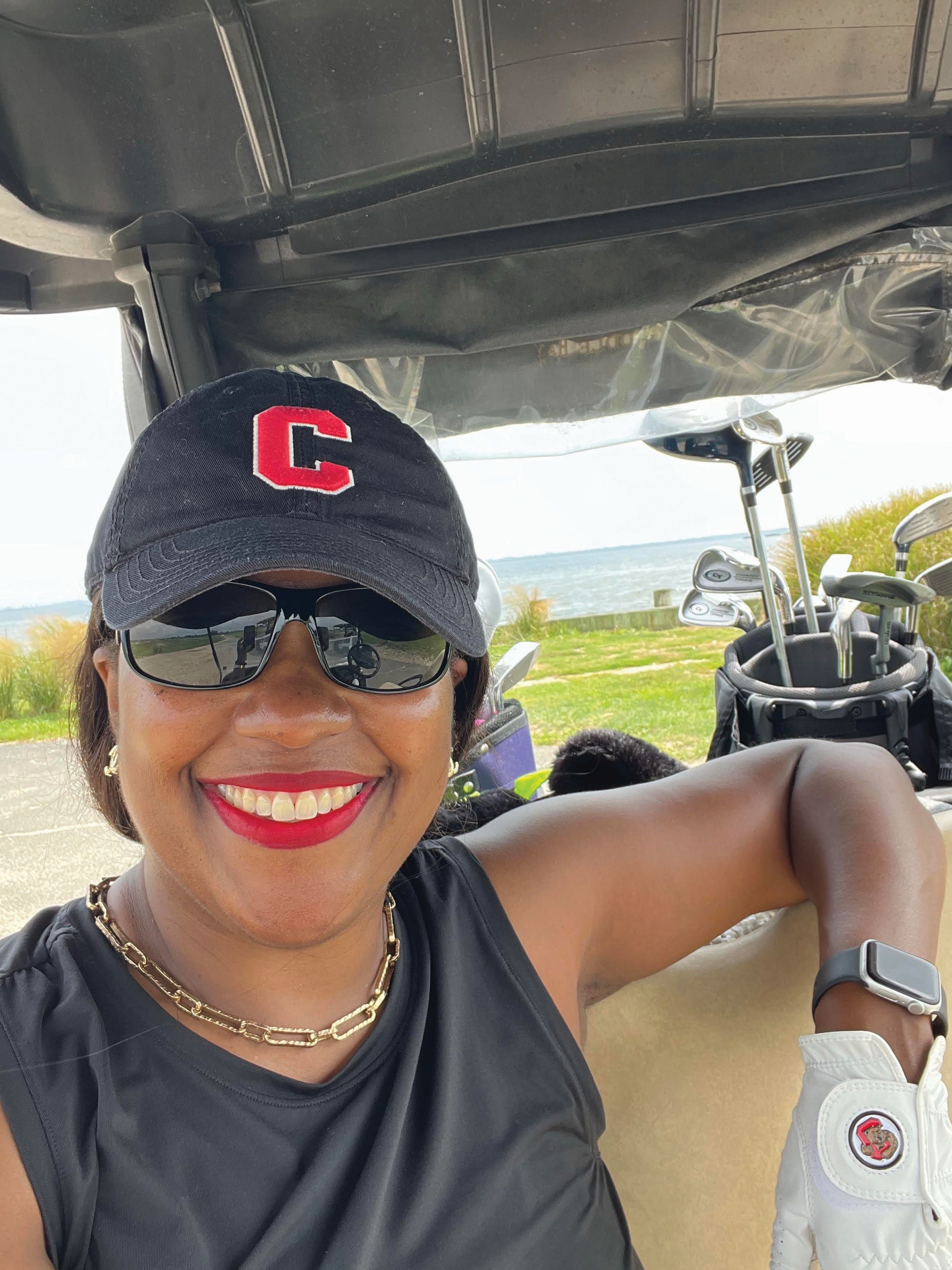
Playing golf
If you could have dinner with any historical figure, who would it be?
Barack Obama
What’s your go-to karaoke song?
Livin’ on a Prayer by Bon Jovi
What’s one thing on your bucket list?
Travel to Rome
If you could instantly learn any skill, what would it be?
Speak Mandarin
What’s your favorite holiday tradition?
Watching Hallmark movies with my daughter
Are you a planner or more spontaneous?
Planner
What’s the best piece of advice you’ve ever received?
Progress, not perfection
Expand Your GI Community with AGA Membership
Renew your AGA membership by Dec. 31 to continue getting to know your AGA Gastro Squad through our monthly column. AGA member spotlights introduce members to one another while recognizing their accomplishments.
Want to recognize a colleague? Submit a nomination to GINews@gastro.org.
Pascale M. White, MD, MBA, MS never tires of excising precancerous polyps.
“To know that I have removed something that could have been potentially dangerous to this patient in years to come, that wasn’t causing any symptoms but silently lurking there” is a great feeling, said Dr. White, an associate professor with dual appointments in the divisions of gastroenterology and liver diseases at the Icahn School of Medicine at Mount Sinai, New York.
“When I do procedures, I always go in with the mindset that this could be a lifesaving procedure for this patient. And that definitely keeps me excited about the field,” she said.
Colorectal cancer is preventable, but when it comes to screening, there are large health disparities. African Americans are 20% more likely to get diagnosed with colorectal cancer and 40% more likely to die from the disease. “Knowing that there are low screening rates among this population, there’s a lot of work to be done with mitigating those disparities,” said Dr. White, who has made it her life’s work to expand access to care and address health inequities.
Dr. White is an inaugural director of Health Equity in Action for Liver and Digestive Diseases (HEALD) and an inaugural fellow of the United Hospital Fund’s Health Equity Fellowship. In 2025, she received the AGA-Pfizer Beacon of Hope Award, which celebrates three women in the GI field who have played a key role in advancing gender and health equity in medicine.
“Through the United Hospital Fund’s Health Equity Fellowship, I have partnered with an East Harlem community health center to conduct seminars and tailor a one-page shared decision tool for colorectal cancer screening to jumpstart discussions on screening choices between patients and providers,” said Dr. White.
In an interview, she offered more details about her mission to connect with communities to improve screening rates for colorectal cancer.
Can you discuss your work with HEALD?
Dr. White: HEALD is a growing initiative to identify and address any access barriers to our screening programs. At this time, I’m working to identify how patients are getting referred to us in our division for colorectal cancer screening and how we can create a more streamlined and robust pathway for patients in the community, namely at federally qualified health centers in East Harlem.
You co-founded the Association of Black Gastroenterologists and Hepatologists (ABGH) in 2021. What are you hoping to accomplish with this organization?
Dr. White: ABGH was co-founded by eleven of us from across the country for the purpose of addressing health care disparities in GI and liver diseases that disproportionately affect Black patient populations. Our mission is to promote health equity, advance science and develop the careers of Black gastroenterologists, hepatologists, and scientists.
Our mentorship program is one way we give back to incoming residents who are interested in pursuing a career in GI. The Nurturing, Excelling, and Unifying Sisters in Medicine (NEXUS) conference centers the perspectives of Black women in medicine from all specialties. The ABGH Summit is an educational conference that features renowned experts in the health equity space.
But at the center of it all is our community outreach. When we started the organization after Chadwick Boseman’s death during the height of the COVID pandemic, all our community events were held over Zoom. Now with our in-person events you can feel the energy in the room. Our main community facing event is called Bustin’ A Gut. It’s a genius combination of comedy and medical education. We have a panel of physicians and comedians. The physicians talk about a range of GI topics such as colorectal cancer screening choices, alarm signs or symptoms of colon cancer, nutrition, and general gut health. The community members feel comfortable asking their questions and the comedians help keep the conversation entertaining and lighthearted. It’s a true laugh and learn event.
How did you become interested in health equity? Was there a specific event or circumstance you could share?
Dr. White: It was my residency training at New York University and my experiences at Bellevue Hospital that really introduced me to a place where everyone could get care. Whether you are coming from another country or right up the street, Bellevue saw everyone who walked through its doors. This is in deep contrast to the vast majority of hospitals where if you do not have insurance, you cannot be seen. Then there are people who have access to care but are overwhelmed by the complexities of the medical system.
Consider colorectal cancer, for example. It is a preventable disease, yet most people aren’t getting screened because they either don’t know they should, they are fearful of the process, or they don’t know how to go about getting the tests done. These are namely knowledge barriers that we can address. I thought: If there’s something I could do to help patients learn about colorectal cancer screening and how they can take steps to prevent this disease, then that’s how I want to spend my career.
You created the Direct Access GI Clinic (DAGIC), one of the projects that led to the AGA-Pfizer Beacon of Hope Award for Gender and Health Equity. How does DAGIC reduce wait times and improve endoscopic care coordination for underserved, high-risk patients?
Dr. White: I developed and implemented a clinic workflow that identified high-risk patients who were sent for direct access procedures but who needed office consultations prior to their procedures. These were the sickest of the sickest patients that needed to be prioritized. Working with my nurse practitioner and office ncurse, we triaged these patients and carved out dedicated time in the week where only DAGIC patients were scheduled.
Creating this direct workflow meant that these patients no longer had to wait three months. They were waiting at most, two to three weeks to be seen. I don’t take for granted that one change in a system can lead to impactful outcomes in patient care and access.
You also co-authored an update to the American College of Gastroenterology’s colorectal cancer screening guidelines for African Americans. Is there anything unique and important that’s worth noting?
Dr. White: We updated those guidelines to include physician recommendation as a potential barrier to screening. We know that patients are more likely to be screened if they are recommended to do so by their physician. Yet, some patients are less likely to receive a physician recommendation for screening. We need to dive deeper into the reasons why this is happening. And if there are any gaps, for example in physician knowledge, that’s something we should readily address.
One of your interests is guiding students, residents, and fellows. What advice would you give to aspiring medical students?
Dr. White: Keep an open mind and explore all your options before committing to a specialty. If you find the field exciting and you are motivated to spend time learning more about it, seek opportunities to conduct research and find a mentor that can further guide you on your journey.
Lightning Round
What’s your favorite season of the year?
Fall
What’s your favorite way to spend a weekend?
Playing golf
If you could have dinner with any historical figure, who would it be?
Barack Obama
What’s your go-to karaoke song?
Livin’ on a Prayer by Bon Jovi
What’s one thing on your bucket list?
Travel to Rome
If you could instantly learn any skill, what would it be?
Speak Mandarin
What’s your favorite holiday tradition?
Watching Hallmark movies with my daughter
Are you a planner or more spontaneous?
Planner
What’s the best piece of advice you’ve ever received?
Progress, not perfection
Expand Your GI Community with AGA Membership
Renew your AGA membership by Dec. 31 to continue getting to know your AGA Gastro Squad through our monthly column. AGA member spotlights introduce members to one another while recognizing their accomplishments.
Want to recognize a colleague? Submit a nomination to GINews@gastro.org.
On a Quest To Reduce Stigmas about Anal Cancer
“I think gastroenterologists are uniquely positioned to help with diagnosing anal diseases, in particular anal cancer,” she said. “It is part of the digestive tract, and my mission is to help gastroenterologists remember that.”
Dr. Korman is a gastroenterologist with Capital Digestive Care in Washington D.C., where she serves as chair of its Women’s Committee and as a member of the board of managers. She’s also the medical director of the Endoscopy Center of Washington D.C.

A recipient of the 2025 AGA Distinguished Clinician Award in Private Practice, Dr. Korman has dedicated her career to educating clinicians on anal cancer screening and anal human papillomavirus. On the research front, she participated as an investigator in the ANAL Cancer-HSIL Outcomes Research (ANCHOR) trial, which led to international anal cancer screening guidelines.
She also co-directs the International Anal Neoplasia Society (IANS) Standard High Resolution Anoscopy course.
When she’s not serving her patients, Dr. Korman speaks in the community about anal cancer awareness and screening. In the last few years, Dr. Korman has presented grand rounds at various institutions and speaks at major medical conferences. “I just try to advocate and help gastroenterologists understand who is at risk, how to look for anal cancer, how to screen, and who to refer. If anyone invites me to speak, I generally will do it,” said Dr. Korman.
In an interview, she talked about the outcomes of the ANCHOR trial and how it may inform future research, and her work to reduce bias and stigma for LGBTQ+ patients.
You decided to become a physician after studying in Egypt and Israel and volunteering with Physicians for Human Rights. Can you talk about that journey?
Dr. Korman: I majored in Religion and Middle East studies, and I minored in Arabic. I thought I was going to become a professor of religious studies. But during my time studying abroad and volunteering for Physicians for Human Rights, I was deeply moved by how physicians connect with the core of our shared humanity. Becoming a physician allows one to meet the most fundamental of human needs—caring for another’s health—in a direct and meaningful way.
My father is a physician, a gastroenterologist, but I never considered it as a career option growing up. The year after I graduated college, I accompanied my parents to my father’s medical school reunion and I thought, ‘Why did I never think about this?’ I decided to go back to school to take the pre-med requirements. Gastroenterology seemed to combine the ability to work with my hands, do procedures, have long-term relationships with patients, and think about complex problems.

GI medicine often involves detective work. What is the most challenging case you’ve encountered?
Dr. Korman: Sometimes the patients who have very severe disorders of gut-brain interaction can be the most challenging because finding treatments for them or getting them to a place where they accept certain types of treatment can be really difficult. And of course, you have to put your detective hat on and make sure you have ruled out all the “zebras.” It can take years to build the level of trust where patients are willing to accept the diagnosis and then pursue appropriate treatment.
I always try my best, but I don’t like to give up. I will refer a patient to a colleague if they have a problem and I can’t figure out what the diagnosis is or find a treatment that works. I believe in second and third opinions. I recognize that there’s a limit to what my brain can do and that we all have blind spots. Maybe someone will look at the case with fresh eyes and think of something else.
What was the most impactful outcomes of the ANAL Cancer-HSIL Outcomes Research (ANCHOR) trial?
Dr. Korman: This was a National Institutes of Health (NIH)-sponsored, randomized controlled trial with 26 clinical sites. We studied people living with human immunodeficiency virus (HIV), as they are the most at-risk group for anal cancer.
We were looking to prove that treating high grade squamous intraepithelial lesions (HSIL) of the anal canal would lead to a significant reduction in the rates of anal cancer. No one in the medical community would accept guidelines or recommendations about what to do with anal pre-cancers until we proved that treatment worked.
We published the findings in 2022. The study concluded when we met our endpoint earlier than expected. We were able to prove that treating high grade anal dysplasia does indeed lead to a very significant reduction in progression to anal cancer. That ultimately led to guidelines. The International Anal Neoplasia Society came out with consensus guidelines on screening for anal cancer in January 2024. In August 2024, NIH, the Centers for Disease Control and Prevention, and the Infectious Diseases Society of America came out with screening guidelines for people living with HIV.
Were there any other outcomes from this research?
Dr. Korman: One of the great things about the study is that we accumulated a bank of tissue and biologic specimens. There were about 4,500 patients randomized into the trial, but about 10,000 patients screened. So, we have a massive collection of biospecimens that we can use to ask questions about the progression of HSIL to anal cancer. We would like to understand more about viral and host molecular mechanisms and hopefully find biomarkers that will identify individuals at particularly high risk of progression. It’s a more precision medicine type of approach.
Education has been a cornerstone of your career. What’s the most rewarding part of teaching the IANS standard high resolution endoscopy course?
Dr. Korman: I first took the course in 2010, and that’s when I started my journey of learning how to perform high resolution endoscopy. Last year I was asked to help co-direct the course. It is now virtual and asynchronous where everything is recorded. But it was exciting to help reorganize the course, update the lectures, and make sure that everything is current. We get to answer questions from participants from all over the world. I think there are participants from 23 countries who have taken the course, which is amazing.
Could you share your work with the LGBTQIA+ population? What specific needs/challenges does this population have with GI care?
Dr. Korman: Many people in the sexual and gender minority community have experienced discrimination in health care settings or know of someone who has. For these reasons, LGBTQIA+ people may approach health care with the expectation of a negative encounter, or they may avoid accessing care altogether. Because anal cancer disproportionately affects sexual and gender minority communities, creating a warm, inclusive environment is key to identifying who is at risk, building trust, and ensuring patients receive the care they need. When you’re talking about anal cancer, there’s a lot of stigma and shame. I think people are afraid to seek care.
Gastroenterology has traditionally been an “old boys club” but that is changing. We’re trying to work on educating people on how to recognize their own biases and move beyond them to provide care that’s affirming and where people feel that they have a safe space to talk about their concerns. Men who have sex with men, in particular living with HIV, are at the highest risk of developing anal cancer. If you don’t know that your patient is a man who has sex with men, or they don’t want to disclose that they’re living with HIV, you don’t know to screen them, and then you’re missing an opportunity to potentially prevent a cancer.

What advice would you give to aspiring medical students interested in GI?
Dr. Korman: GI is the most exciting and interesting field. We take care of so many different organs, and we’re never bored. If medical students want to get into GI, I recommend that they try to be in an office or an endoscopy center and see if it’s really for them and get some hands-on experience if possible. To be truly great at this profession, you really must see it as a calling – jump in with your whole heart and not see it as just a job. If you can do that, you’ll succeed.
How do you handle stress and maintain work-life balance?
Dr. Korman: Exercise. I try to work out at least five days a week. I can’t live without it. That keeps me going. What do I do for fun? I spend time with my family and my friends. I enjoy going to new restaurants and being outdoors, especially near a body of water. I travel, and I love watching movies. I am also guilty of binge-watching TV on a regular basis as well.
Lightning Round
Coffee or tea?
Coffee, 100%
What’s your favorite book?
I can’t say I have just one, but I recently read Tomorrow and Tomorrow and Tomorrow and loved it
Beach vacation or mountain retreat?
Beach
Early bird or night owl?
Early bird
What’s your go-to comfort food?
Anything with bananas
If you could travel anywhere, where would you go?
Vietnam or African safari
What’s your favorite childhood memory?
Swim team when I was a kid
If you could instantly learn any skill, what would it be?
Playing the drums
Are you a planner or more spontaneous?
Planner, although it’s not my strong suit, if I’m being honest.
“I think gastroenterologists are uniquely positioned to help with diagnosing anal diseases, in particular anal cancer,” she said. “It is part of the digestive tract, and my mission is to help gastroenterologists remember that.”
Dr. Korman is a gastroenterologist with Capital Digestive Care in Washington D.C., where she serves as chair of its Women’s Committee and as a member of the board of managers. She’s also the medical director of the Endoscopy Center of Washington D.C.
A recipient of the 2025 AGA Distinguished Clinician Award in Private Practice, Dr. Korman has dedicated her career to educating clinicians on anal cancer screening and anal human papillomavirus. On the research front, she participated as an investigator in the ANAL Cancer-HSIL Outcomes Research (ANCHOR) trial, which led to international anal cancer screening guidelines.
She also co-directs the International Anal Neoplasia Society (IANS) Standard High Resolution Anoscopy course.
When she’s not serving her patients, Dr. Korman speaks in the community about anal cancer awareness and screening. In the last few years, Dr. Korman has presented grand rounds at various institutions and speaks at major medical conferences. “I just try to advocate and help gastroenterologists understand who is at risk, how to look for anal cancer, how to screen, and who to refer. If anyone invites me to speak, I generally will do it,” said Dr. Korman.
In an interview, she talked about the outcomes of the ANCHOR trial and how it may inform future research, and her work to reduce bias and stigma for LGBTQ+ patients.
You decided to become a physician after studying in Egypt and Israel and volunteering with Physicians for Human Rights. Can you talk about that journey?
Dr. Korman: I majored in Religion and Middle East studies, and I minored in Arabic. I thought I was going to become a professor of religious studies. But during my time studying abroad and volunteering for Physicians for Human Rights, I was deeply moved by how physicians connect with the core of our shared humanity. Becoming a physician allows one to meet the most fundamental of human needs—caring for another’s health—in a direct and meaningful way.
My father is a physician, a gastroenterologist, but I never considered it as a career option growing up. The year after I graduated college, I accompanied my parents to my father’s medical school reunion and I thought, ‘Why did I never think about this?’ I decided to go back to school to take the pre-med requirements. Gastroenterology seemed to combine the ability to work with my hands, do procedures, have long-term relationships with patients, and think about complex problems.
GI medicine often involves detective work. What is the most challenging case you’ve encountered?
Dr. Korman: Sometimes the patients who have very severe disorders of gut-brain interaction can be the most challenging because finding treatments for them or getting them to a place where they accept certain types of treatment can be really difficult. And of course, you have to put your detective hat on and make sure you have ruled out all the “zebras.” It can take years to build the level of trust where patients are willing to accept the diagnosis and then pursue appropriate treatment.
I always try my best, but I don’t like to give up. I will refer a patient to a colleague if they have a problem and I can’t figure out what the diagnosis is or find a treatment that works. I believe in second and third opinions. I recognize that there’s a limit to what my brain can do and that we all have blind spots. Maybe someone will look at the case with fresh eyes and think of something else.
What was the most impactful outcomes of the ANAL Cancer-HSIL Outcomes Research (ANCHOR) trial?
Dr. Korman: This was a National Institutes of Health (NIH)-sponsored, randomized controlled trial with 26 clinical sites. We studied people living with human immunodeficiency virus (HIV), as they are the most at-risk group for anal cancer.
We were looking to prove that treating high grade squamous intraepithelial lesions (HSIL) of the anal canal would lead to a significant reduction in the rates of anal cancer. No one in the medical community would accept guidelines or recommendations about what to do with anal pre-cancers until we proved that treatment worked.
We published the findings in 2022. The study concluded when we met our endpoint earlier than expected. We were able to prove that treating high grade anal dysplasia does indeed lead to a very significant reduction in progression to anal cancer. That ultimately led to guidelines. The International Anal Neoplasia Society came out with consensus guidelines on screening for anal cancer in January 2024. In August 2024, NIH, the Centers for Disease Control and Prevention, and the Infectious Diseases Society of America came out with screening guidelines for people living with HIV.
Were there any other outcomes from this research?
Dr. Korman: One of the great things about the study is that we accumulated a bank of tissue and biologic specimens. There were about 4,500 patients randomized into the trial, but about 10,000 patients screened. So, we have a massive collection of biospecimens that we can use to ask questions about the progression of HSIL to anal cancer. We would like to understand more about viral and host molecular mechanisms and hopefully find biomarkers that will identify individuals at particularly high risk of progression. It’s a more precision medicine type of approach.
Education has been a cornerstone of your career. What’s the most rewarding part of teaching the IANS standard high resolution endoscopy course?
Dr. Korman: I first took the course in 2010, and that’s when I started my journey of learning how to perform high resolution endoscopy. Last year I was asked to help co-direct the course. It is now virtual and asynchronous where everything is recorded. But it was exciting to help reorganize the course, update the lectures, and make sure that everything is current. We get to answer questions from participants from all over the world. I think there are participants from 23 countries who have taken the course, which is amazing.
Could you share your work with the LGBTQIA+ population? What specific needs/challenges does this population have with GI care?
Dr. Korman: Many people in the sexual and gender minority community have experienced discrimination in health care settings or know of someone who has. For these reasons, LGBTQIA+ people may approach health care with the expectation of a negative encounter, or they may avoid accessing care altogether. Because anal cancer disproportionately affects sexual and gender minority communities, creating a warm, inclusive environment is key to identifying who is at risk, building trust, and ensuring patients receive the care they need. When you’re talking about anal cancer, there’s a lot of stigma and shame. I think people are afraid to seek care.
Gastroenterology has traditionally been an “old boys club” but that is changing. We’re trying to work on educating people on how to recognize their own biases and move beyond them to provide care that’s affirming and where people feel that they have a safe space to talk about their concerns. Men who have sex with men, in particular living with HIV, are at the highest risk of developing anal cancer. If you don’t know that your patient is a man who has sex with men, or they don’t want to disclose that they’re living with HIV, you don’t know to screen them, and then you’re missing an opportunity to potentially prevent a cancer.
What advice would you give to aspiring medical students interested in GI?
Dr. Korman: GI is the most exciting and interesting field. We take care of so many different organs, and we’re never bored. If medical students want to get into GI, I recommend that they try to be in an office or an endoscopy center and see if it’s really for them and get some hands-on experience if possible. To be truly great at this profession, you really must see it as a calling – jump in with your whole heart and not see it as just a job. If you can do that, you’ll succeed.
How do you handle stress and maintain work-life balance?
Dr. Korman: Exercise. I try to work out at least five days a week. I can’t live without it. That keeps me going. What do I do for fun? I spend time with my family and my friends. I enjoy going to new restaurants and being outdoors, especially near a body of water. I travel, and I love watching movies. I am also guilty of binge-watching TV on a regular basis as well.
Lightning Round
Coffee or tea?
Coffee, 100%
What’s your favorite book?
I can’t say I have just one, but I recently read Tomorrow and Tomorrow and Tomorrow and loved it
Beach vacation or mountain retreat?
Beach
Early bird or night owl?
Early bird
What’s your go-to comfort food?
Anything with bananas
If you could travel anywhere, where would you go?
Vietnam or African safari
What’s your favorite childhood memory?
Swim team when I was a kid
If you could instantly learn any skill, what would it be?
Playing the drums
Are you a planner or more spontaneous?
Planner, although it’s not my strong suit, if I’m being honest.

Bringing HCC Patients Hope Through Trials, Advanced Treatments
For Reena Salgia, MD, the most rewarding part about working with patients with hepatocellular carcinoma is being there for their entire journey, thanks to advancements in treatment. “It brings a smile to my face just to think about it,” says Dr. Salgia, medical director of Henry Ford Health’s Liver Cancer Clinic in Detroit.
Hepatocellular carcinoma accounts for 80% of all liver cancer. When she first entered the field, Dr. Salgia often heard that survival rates 5 years after diagnosis were less than 10%. Over the last decade however, “I’ve seen an expansion in the procedural options that we offer these patients. We have an array of options both surgically as well as procedurally,” she said.
Especially over the last three to four years, “we’ve seen meaningful responses for patients with medications that we previously didn’t have in our toolbox. That’s really been exciting, along with continued involvement in clinical trials and being able to offer patients a number of different approaches to their care of liver cancer,” said Dr. Salgia.
A regular attendee and presenter at national GI meetings, Dr. Salgia participated in AGA’s Women’s Executive Leadership Conference in 2023. Her academic resume includes a long list of clinical trials to assess treatments for patients at different stages of hepatocellular carcinoma.
In an interview, she discussed the highlights of her career as a researcher and mentor of fellows, and how she guides and supports her transplant patients.
What drove you to pursue the field of hepatology and transplant hepatology?
I came across this field during my fourth year of medical school. I didn’t know anything about hepatology when I reached that stage and had the opportunity to do an elective. I just fell in love with the specialty. I liked the complex pathophysiology of liver disease, the long-term follow-up and care of patients. It appealed to the type of science that I had enjoyed back in college.
As I went into my GI fellowship training, I got to learn more about the field of transplant medicine. For instance, how you can take these patients who are incredibly ill, really at a very vulnerable point of their illness, and then offer them great hope and see their lives turn around afterwards. When I had the opportunity to see patients go from end stage liver disease to such significant improvement in their quality of life, and restoring their physical functioning beyond what we would’ve ever imagined when they were ill, it reaffirmed my interest in both hepatology as well as in transplant medicine.
How do you help those patients waiting on transplant lists for a liver?
We are intimately involved in their care all the way through their journey with liver disease, up until the time of physically getting the liver transplant, which is performed by our colleagues in transplant surgery. From the time they are transplanted, we are involved in their inpatient and outpatient post-transplant care. We’ve helped to get them on the transplant list with the work of the multidisciplinary team. If there are opportunities to help them understand their position on the list or obtaining exceptions—though that is done in a very objective fashion through the regulatory system—we help to guide them through that journey.
You’ve worked on many studies that involve treatments for hepatocellular carcinoma. Can you highlight a paper that yielded clinically significant benefits?
What really stands out the most to me was our site’s involvement in the IMbrave150 trial, which was published in 2020. This multicenter study made a big difference in the outcomes and treatments for patients, as it brought the adoption of first-line immunotherapy (atezolizumab plus bevacizumab) for patients with advanced hepatocellular carcinoma. I remember vividly the patients we had the opportunity to enroll in that trial – some who we continue to care for today. This stands out as one of the trials that I was involved in that had a lasting impact.
What were the clinical endpoints and key results of that trial?
The endpoint was to see an improvement in overall survival utilizing immunotherapy, compared with the prior standard of care then available, oral therapy. The results led to the adoption and FDA approval of immunotherapy in the first line setting for advanced unresectable hepatocellular carcinoma patients.
What are some of the highlights of serving as director of Henry Ford’s fellowship program?
Education is my passion. I went into medical training feeling that at some point I would love to blend in teaching in a formal role. Becoming program director of the gastroenterology and hepatology fellowship at Henry Ford in 2018 was one of the most meaningful things that I’ve had the opportunity to do in my career. I get to see trainees who are at a very impressionable point of their journey go on to become gastroenterologists and then launch into their first job and really develop in this field. Seeing them come in day one, not knowing how to hold a scope or do a procedure on a patient of this nature, then quickly evolve over the first year and grow over three years to achieve this specialty training [is rewarding]. I’ve learned a lot from the fellows along the way. I think of them as an extension of my family. We have 15 fellows currently in our program and we’ll be growing this summer. So that’s really been a highlight of my career thus far.
What fears did you have to push past to get to where you are in your career?
I think that there have been a few. One is certainly the fear of making the wrong choice with your first career opportunity. I did choose to leave my comfort zone from where I had done my training. I met that with some fear, but also excitement for new opportunities of personal and professional growth.
Another fear is: Am I going to be able to be ambitious in this field? Can I pursue research, become a program director, and do things that my role models and mentors were able to achieve? There’s also the fear of being able to balance a busy work life with a busy home life and figuring out how to do both well and minimize the guilt on both sides. I have a family with two girls. They are definitely a top priority.
What teacher or mentor had the greatest impact on you?
Helen Te, MD, a hepatologist at the University of Chicago. When I was a medical student there, I had the opportunity to work with her and saw her passion for this field. She really had so much enthusiasm for teaching and was a big part of why I started to fall in love with liver disease.

Karen Kim, MD, now the dean of Penn State College of Medicine, was one of my assigned mentors as a medical student. She helped me explore the fields where there were opportunities for residency and helped me make the decision to go into internal medicine, which often is a key deciding point for medical students. She was also a very influential teacher. The other individual who stands out is my fellowship program director, Hari Sree Conjeevaram, MD, MSc, at University of Michigan Health. He exhibited the qualities as an educator and program director that helped me recognize that education was something that I wanted to pursue in a formal fashion once I moved on in my career.
Describe how you would spend a free Saturday afternoon.
Likely taking a hike or go to a park with my family, enjoying the outdoors and spending time with them.
Lightning Round
If you weren’t a gastroenterologist, what would you be?
Philanthropist
Favorite city in U.S. besides the one you live in?
Chicago
Place you most want to travel?
New Zealand
Favorite breakfast?
Avocado toast
Favorite ice cream flavor?
Cookies and cream
How many cups of coffee do you drink per day?
Two…or more
Cat person or dog person?
Dog
Texting or talking?
Talk
Favorite season?
Autumn
Favorite type of music?
Pop
Favorite movie genre?
Action
For Reena Salgia, MD, the most rewarding part about working with patients with hepatocellular carcinoma is being there for their entire journey, thanks to advancements in treatment. “It brings a smile to my face just to think about it,” says Dr. Salgia, medical director of Henry Ford Health’s Liver Cancer Clinic in Detroit.
Hepatocellular carcinoma accounts for 80% of all liver cancer. When she first entered the field, Dr. Salgia often heard that survival rates 5 years after diagnosis were less than 10%. Over the last decade however, “I’ve seen an expansion in the procedural options that we offer these patients. We have an array of options both surgically as well as procedurally,” she said.
Especially over the last three to four years, “we’ve seen meaningful responses for patients with medications that we previously didn’t have in our toolbox. That’s really been exciting, along with continued involvement in clinical trials and being able to offer patients a number of different approaches to their care of liver cancer,” said Dr. Salgia.
A regular attendee and presenter at national GI meetings, Dr. Salgia participated in AGA’s Women’s Executive Leadership Conference in 2023. Her academic resume includes a long list of clinical trials to assess treatments for patients at different stages of hepatocellular carcinoma.
In an interview, she discussed the highlights of her career as a researcher and mentor of fellows, and how she guides and supports her transplant patients.
What drove you to pursue the field of hepatology and transplant hepatology?
I came across this field during my fourth year of medical school. I didn’t know anything about hepatology when I reached that stage and had the opportunity to do an elective. I just fell in love with the specialty. I liked the complex pathophysiology of liver disease, the long-term follow-up and care of patients. It appealed to the type of science that I had enjoyed back in college.
As I went into my GI fellowship training, I got to learn more about the field of transplant medicine. For instance, how you can take these patients who are incredibly ill, really at a very vulnerable point of their illness, and then offer them great hope and see their lives turn around afterwards. When I had the opportunity to see patients go from end stage liver disease to such significant improvement in their quality of life, and restoring their physical functioning beyond what we would’ve ever imagined when they were ill, it reaffirmed my interest in both hepatology as well as in transplant medicine.
How do you help those patients waiting on transplant lists for a liver?
We are intimately involved in their care all the way through their journey with liver disease, up until the time of physically getting the liver transplant, which is performed by our colleagues in transplant surgery. From the time they are transplanted, we are involved in their inpatient and outpatient post-transplant care. We’ve helped to get them on the transplant list with the work of the multidisciplinary team. If there are opportunities to help them understand their position on the list or obtaining exceptions—though that is done in a very objective fashion through the regulatory system—we help to guide them through that journey.
You’ve worked on many studies that involve treatments for hepatocellular carcinoma. Can you highlight a paper that yielded clinically significant benefits?
What really stands out the most to me was our site’s involvement in the IMbrave150 trial, which was published in 2020. This multicenter study made a big difference in the outcomes and treatments for patients, as it brought the adoption of first-line immunotherapy (atezolizumab plus bevacizumab) for patients with advanced hepatocellular carcinoma. I remember vividly the patients we had the opportunity to enroll in that trial – some who we continue to care for today. This stands out as one of the trials that I was involved in that had a lasting impact.
What were the clinical endpoints and key results of that trial?
The endpoint was to see an improvement in overall survival utilizing immunotherapy, compared with the prior standard of care then available, oral therapy. The results led to the adoption and FDA approval of immunotherapy in the first line setting for advanced unresectable hepatocellular carcinoma patients.
What are some of the highlights of serving as director of Henry Ford’s fellowship program?
Education is my passion. I went into medical training feeling that at some point I would love to blend in teaching in a formal role. Becoming program director of the gastroenterology and hepatology fellowship at Henry Ford in 2018 was one of the most meaningful things that I’ve had the opportunity to do in my career. I get to see trainees who are at a very impressionable point of their journey go on to become gastroenterologists and then launch into their first job and really develop in this field. Seeing them come in day one, not knowing how to hold a scope or do a procedure on a patient of this nature, then quickly evolve over the first year and grow over three years to achieve this specialty training [is rewarding]. I’ve learned a lot from the fellows along the way. I think of them as an extension of my family. We have 15 fellows currently in our program and we’ll be growing this summer. So that’s really been a highlight of my career thus far.
What fears did you have to push past to get to where you are in your career?
I think that there have been a few. One is certainly the fear of making the wrong choice with your first career opportunity. I did choose to leave my comfort zone from where I had done my training. I met that with some fear, but also excitement for new opportunities of personal and professional growth.
Another fear is: Am I going to be able to be ambitious in this field? Can I pursue research, become a program director, and do things that my role models and mentors were able to achieve? There’s also the fear of being able to balance a busy work life with a busy home life and figuring out how to do both well and minimize the guilt on both sides. I have a family with two girls. They are definitely a top priority.
What teacher or mentor had the greatest impact on you?
Helen Te, MD, a hepatologist at the University of Chicago. When I was a medical student there, I had the opportunity to work with her and saw her passion for this field. She really had so much enthusiasm for teaching and was a big part of why I started to fall in love with liver disease.
Karen Kim, MD, now the dean of Penn State College of Medicine, was one of my assigned mentors as a medical student. She helped me explore the fields where there were opportunities for residency and helped me make the decision to go into internal medicine, which often is a key deciding point for medical students. She was also a very influential teacher. The other individual who stands out is my fellowship program director, Hari Sree Conjeevaram, MD, MSc, at University of Michigan Health. He exhibited the qualities as an educator and program director that helped me recognize that education was something that I wanted to pursue in a formal fashion once I moved on in my career.
Describe how you would spend a free Saturday afternoon.
Likely taking a hike or go to a park with my family, enjoying the outdoors and spending time with them.
Lightning Round
If you weren’t a gastroenterologist, what would you be?
Philanthropist
Favorite city in U.S. besides the one you live in?
Chicago
Place you most want to travel?
New Zealand
Favorite breakfast?
Avocado toast
Favorite ice cream flavor?
Cookies and cream
How many cups of coffee do you drink per day?
Two…or more
Cat person or dog person?
Dog
Texting or talking?
Talk
Favorite season?
Autumn
Favorite type of music?
Pop
Favorite movie genre?
Action
Endoscopist Brings Cutting-Edge Tech to Asia-Pacific Region
As the COVID-19 crisis unfolded in early 2020, Tossapol Kerdsirichairat, MD, faced another challenge: his mother’s ovarian cancer diagnosis.
“She chose to remain in Thailand, so I decided to relocate to care for her,” said Dr. Kerdsirichairat, an interventional endoscopist who completed fellowships at the University of Michigan, Ann Arbor, and Johns Hopkins University in Baltimore. The move to Bangkok turned out to be one of the best decisions of his life, he said, as he could support his mother while introducing advanced endoscopic techniques and devices to the region.
“Bangkok is a hub for medical innovation in Asia, offering opportunities to work with a diverse patient population and access to cutting-edge technology,” said Dr. Kerdsirichairat, who works at Bumrungrad International Hospital as a clinical associate professor.
The program is the first of its kind in Thailand and one of the few in the Asia-Pacific region.
“I guide patients and families through understanding their risks and implementing preventive strategies, collaborating with multidisciplinary teams to ensure comprehensive care. It’s incredibly rewarding to see the impact of early tumor detection,” said Dr. Kerdsirichairat, an international member of AGA who was a participant in the AGA Young Delegates Program.
He has set several records in Thailand for the smallest tumor detected, including a 0.3-millimeter (mm) esophageal tumor, a 0.8-mm tumor for stomach cancer, a 5-mm pancreatic tumor, and a 1-mm tumor for colon cancer.
“These were detected through high-standard screening programs, as patients often do not develop symptoms from these subtle lesions,” said Dr. Kerdsirichairat, who discussed in an interview the unique challenges of practicing overseas.
Why did you choose GI?
Gastroenterology is a specialty that uniquely integrates procedural skill, clinical decision making, and a deep understanding of complex biological systems. I was drawn especially to the ability to make a direct and meaningful impact in patients’ lives through advanced endoscopic procedures, while also addressing both acute and chronic diseases, and focusing on cancer prevention. It is incredibly rewarding to perform an endoscopic retrograde cholangiopancreatography (ERCP) for cholangitis and see a patient return to normal the very next day, or to perform an endoscopic ultrasound (EUS) for pancreatic cancer screening in high-risk individuals and detect a sub-centimeter pancreatic tumor.
Realizing that early detection can improve survival by threefold after surgery is a powerful reminder of the difference we can make in patients’ lives. This specialty requires a delicate balance of precision and empathy, which perfectly aligns with my strengths and values as a physician.
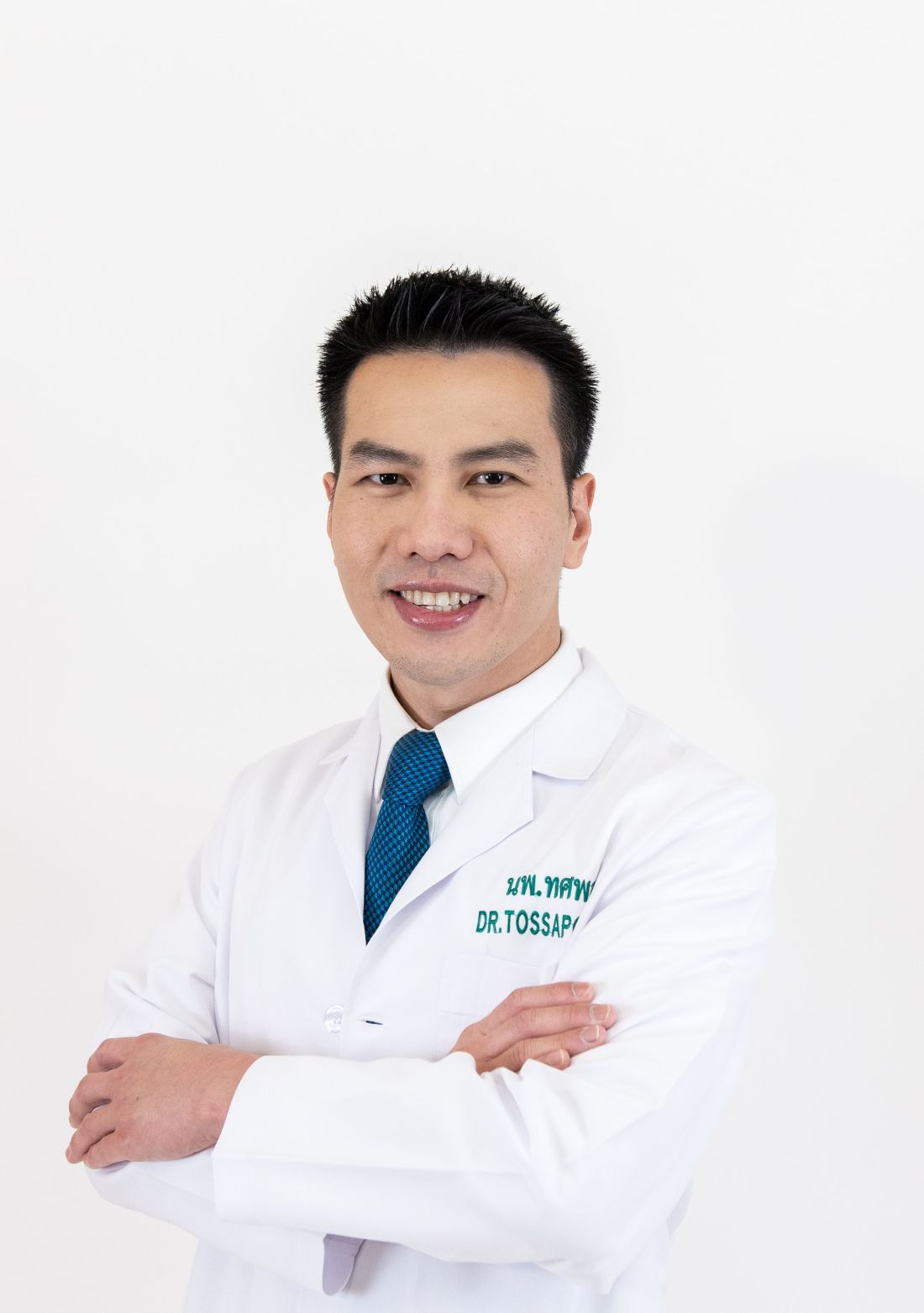
You have a wide variety of clinical interests, from endoscopic procedures to cancer research to GERD. What’s your key subspecialty and why?
My primary specialty is advanced endoscopy, which includes techniques such as EUS, ERCP, and endoscopic resection of precancerous and early cancerous lesions. I also focus on cutting-edge, evidence-based techniques recently included in clinical guidelines, such as Transoral Incisionless Fundoplication (TIF). These minimally invasive options allow me to diagnose and treat conditions that once required surgery. The precision and innovation involved in advanced endoscopy enable me to effectively manage complex cases—from diagnosing early cancers to managing bile duct obstructions and resecting precancerous lesions.
Can you describe your work in cancer genetics and screening?
I am deeply committed to the early detection of gastrointestinal cancers, particularly through screening for precancerous conditions and hereditary syndromes. During my general GI training at the University of Michigan, I had the privilege of working with Grace Elta, MD, AGAF, and Michelle Anderson, MD, MSc, renowned experts in pancreatic cancer management. I was later trained by Anne Marie Lennon, PhD, AGAF, who pioneered the liquid biopsy technique for cancer screening through the CancerSEEK project, and Marcia (Mimi) Canto, MD, MHS, who initiated the Cancer of the Pancreas Screening project for high-risk individuals of pancreatic cancer.
I also had the distinction of being the first at Bumrungrad International Hospital to perform endoscopic drainage for pancreatic fluid collections in the setting of multi-organ failure. This endoscopic approach has been extensively validated in the medical literature as significantly improving survival rates compared to surgical drainage. My training in this specialized procedure was conducted under the guidance of the premier group for necrotizing pancreatitis, led by Martin Freeman, MD, at the University of Minnesota.
Later, I contributed to overseeing the Inherited Gastrointestinal Malignancy Clinic of MyCode, a large-scale population-based cohort program focused on cancer screening in Pennsylvania. By December 2024, MyCode had collected blood samples from over 258,000 individuals, analyzed DNA sequences from over 184,000, and provided clinical data that benefits over 142,000 patients. It’s not uncommon for healthy 25-year-old patients to come to our clinic for colon cancer screening after learning from the program that they carry a cancer syndrome, and early screening can potentially save their lives.
What are the key differences between training and practicing medicine in the United States and in an Asian country?
The U.S. healthcare system is deeply rooted in evidence-based protocols and multidisciplinary care, driven by an insurance-based model. In contrast, many Asian countries face challenges such as the dependency on government approval for certain treatments and insurance limitations. Practicing in Asia requires navigating unique cultural, economic, and systemic differences, including varying resource availability and disease prevalence.
What specific challenges have you faced as a GI in Thailand?
As an advanced endoscopist, one of the biggest challenges I faced initially was the difficulty in obtaining the same devices I used in the U.S. for use in Thailand. With support from device companies and mentors in the U.S., I was able to perform groundbreaking procedures, such as the TIF in Southeast Asia and the first use of a full-thickness resection device in Thailand. I am also proud to be part of one of the first few centers worldwide performing the combination of injectable semaglutide and endoscopic sleeve gastroplasty, resulting in a remarkable weight reduction of 44%, comparable to surgical gastric bypass.
In addition, Bumrungrad International Hospital, where I practice, sees over 1.1 million visits annually from patients from more than 190 countries. This offers a unique opportunity to engage with a global patient base and learn from diverse cultures. Over time, although the hospital has professional interpreters for all languages, I have become able to communicate basic sentences with international patients in their preferred languages, including Chinese, Japanese, and Arabic, which has enriched my practice.
What’s your favorite thing to do when you’re not practicing GI?
I enjoy traveling, exploring new cuisines, and spending quality time with family and friends. These activities help me recharge and offer fresh perspectives on life.
Lightning Round
Texting or talking?
Talking. It’s more personal and meaningful.
Favorite city in the U.S.?
Ann Arbor, Michigan
Cat or dog person?
Dog person
Favorite junk food?
Pizza
How many cups of coffee do you drink per day?
Two – just enough to stay sharp, but not jittery.
If you weren’t a GI, what would you be?
Architect
Best place you went on vacation?
Kyoto, Japan
Favorite sport?
Skiing
Favorite ice cream?
Matcha green tea
What song do you have to sing along with when you hear it?
“Everybody” by Backstreet Boys
Favorite movie or TV show?
Forrest Gump and Friends
Optimist or pessimist?
Optimist. I believe in focusing on solutions and possibilities.
As the COVID-19 crisis unfolded in early 2020, Tossapol Kerdsirichairat, MD, faced another challenge: his mother’s ovarian cancer diagnosis.
“She chose to remain in Thailand, so I decided to relocate to care for her,” said Dr. Kerdsirichairat, an interventional endoscopist who completed fellowships at the University of Michigan, Ann Arbor, and Johns Hopkins University in Baltimore. The move to Bangkok turned out to be one of the best decisions of his life, he said, as he could support his mother while introducing advanced endoscopic techniques and devices to the region.
“Bangkok is a hub for medical innovation in Asia, offering opportunities to work with a diverse patient population and access to cutting-edge technology,” said Dr. Kerdsirichairat, who works at Bumrungrad International Hospital as a clinical associate professor.
The program is the first of its kind in Thailand and one of the few in the Asia-Pacific region.
“I guide patients and families through understanding their risks and implementing preventive strategies, collaborating with multidisciplinary teams to ensure comprehensive care. It’s incredibly rewarding to see the impact of early tumor detection,” said Dr. Kerdsirichairat, an international member of AGA who was a participant in the AGA Young Delegates Program.
He has set several records in Thailand for the smallest tumor detected, including a 0.3-millimeter (mm) esophageal tumor, a 0.8-mm tumor for stomach cancer, a 5-mm pancreatic tumor, and a 1-mm tumor for colon cancer.
“These were detected through high-standard screening programs, as patients often do not develop symptoms from these subtle lesions,” said Dr. Kerdsirichairat, who discussed in an interview the unique challenges of practicing overseas.
Why did you choose GI?
Gastroenterology is a specialty that uniquely integrates procedural skill, clinical decision making, and a deep understanding of complex biological systems. I was drawn especially to the ability to make a direct and meaningful impact in patients’ lives through advanced endoscopic procedures, while also addressing both acute and chronic diseases, and focusing on cancer prevention. It is incredibly rewarding to perform an endoscopic retrograde cholangiopancreatography (ERCP) for cholangitis and see a patient return to normal the very next day, or to perform an endoscopic ultrasound (EUS) for pancreatic cancer screening in high-risk individuals and detect a sub-centimeter pancreatic tumor.
Realizing that early detection can improve survival by threefold after surgery is a powerful reminder of the difference we can make in patients’ lives. This specialty requires a delicate balance of precision and empathy, which perfectly aligns with my strengths and values as a physician.
You have a wide variety of clinical interests, from endoscopic procedures to cancer research to GERD. What’s your key subspecialty and why?
My primary specialty is advanced endoscopy, which includes techniques such as EUS, ERCP, and endoscopic resection of precancerous and early cancerous lesions. I also focus on cutting-edge, evidence-based techniques recently included in clinical guidelines, such as Transoral Incisionless Fundoplication (TIF). These minimally invasive options allow me to diagnose and treat conditions that once required surgery. The precision and innovation involved in advanced endoscopy enable me to effectively manage complex cases—from diagnosing early cancers to managing bile duct obstructions and resecting precancerous lesions.
Can you describe your work in cancer genetics and screening?
I am deeply committed to the early detection of gastrointestinal cancers, particularly through screening for precancerous conditions and hereditary syndromes. During my general GI training at the University of Michigan, I had the privilege of working with Grace Elta, MD, AGAF, and Michelle Anderson, MD, MSc, renowned experts in pancreatic cancer management. I was later trained by Anne Marie Lennon, PhD, AGAF, who pioneered the liquid biopsy technique for cancer screening through the CancerSEEK project, and Marcia (Mimi) Canto, MD, MHS, who initiated the Cancer of the Pancreas Screening project for high-risk individuals of pancreatic cancer.
I also had the distinction of being the first at Bumrungrad International Hospital to perform endoscopic drainage for pancreatic fluid collections in the setting of multi-organ failure. This endoscopic approach has been extensively validated in the medical literature as significantly improving survival rates compared to surgical drainage. My training in this specialized procedure was conducted under the guidance of the premier group for necrotizing pancreatitis, led by Martin Freeman, MD, at the University of Minnesota.
Later, I contributed to overseeing the Inherited Gastrointestinal Malignancy Clinic of MyCode, a large-scale population-based cohort program focused on cancer screening in Pennsylvania. By December 2024, MyCode had collected blood samples from over 258,000 individuals, analyzed DNA sequences from over 184,000, and provided clinical data that benefits over 142,000 patients. It’s not uncommon for healthy 25-year-old patients to come to our clinic for colon cancer screening after learning from the program that they carry a cancer syndrome, and early screening can potentially save their lives.
What are the key differences between training and practicing medicine in the United States and in an Asian country?
The U.S. healthcare system is deeply rooted in evidence-based protocols and multidisciplinary care, driven by an insurance-based model. In contrast, many Asian countries face challenges such as the dependency on government approval for certain treatments and insurance limitations. Practicing in Asia requires navigating unique cultural, economic, and systemic differences, including varying resource availability and disease prevalence.
What specific challenges have you faced as a GI in Thailand?
As an advanced endoscopist, one of the biggest challenges I faced initially was the difficulty in obtaining the same devices I used in the U.S. for use in Thailand. With support from device companies and mentors in the U.S., I was able to perform groundbreaking procedures, such as the TIF in Southeast Asia and the first use of a full-thickness resection device in Thailand. I am also proud to be part of one of the first few centers worldwide performing the combination of injectable semaglutide and endoscopic sleeve gastroplasty, resulting in a remarkable weight reduction of 44%, comparable to surgical gastric bypass.
In addition, Bumrungrad International Hospital, where I practice, sees over 1.1 million visits annually from patients from more than 190 countries. This offers a unique opportunity to engage with a global patient base and learn from diverse cultures. Over time, although the hospital has professional interpreters for all languages, I have become able to communicate basic sentences with international patients in their preferred languages, including Chinese, Japanese, and Arabic, which has enriched my practice.
What’s your favorite thing to do when you’re not practicing GI?
I enjoy traveling, exploring new cuisines, and spending quality time with family and friends. These activities help me recharge and offer fresh perspectives on life.
Lightning Round
Texting or talking?
Talking. It’s more personal and meaningful.
Favorite city in the U.S.?
Ann Arbor, Michigan
Cat or dog person?
Dog person
Favorite junk food?
Pizza
How many cups of coffee do you drink per day?
Two – just enough to stay sharp, but not jittery.
If you weren’t a GI, what would you be?
Architect
Best place you went on vacation?
Kyoto, Japan
Favorite sport?
Skiing
Favorite ice cream?
Matcha green tea
What song do you have to sing along with when you hear it?
“Everybody” by Backstreet Boys
Favorite movie or TV show?
Forrest Gump and Friends
Optimist or pessimist?
Optimist. I believe in focusing on solutions and possibilities.
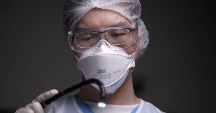
Three Sisters Embrace ‘Collaborative Spirit’ of GI Science
They all share the same genes—and job title.
Each has her own lab, working in different specialties. But if one sister needs the others, it’s reassuring to know they’re not far away.
“We have very different points of view. I’m interested in microbes. Amy’s really interested in myosin mediated trafficking and Kristen’s interested in viruses and purinergic signaling. It’s awesome that we can all work in the same field but have very different questions. And there’s so many questions that we can tackle,” said Mindy Engevik, the oldest of the trio.

If Mindy’s students need help with staining, she sends them to Amy’s lab. If they need help with calcium signaling and live cell imaging, she’ll send them to Kristen’s lab. “We interchange our expertise a lot,” said Mindy.
It’s nice to have a sister down the hall at work who can advise you on RNA sequencing analysis or immunofluorescence imaging, noted Amy Engevik. “You can ask them: ‘Can you just walk my student through this for a minute?’ Or, could they help with organoid cultures you don’t have time for right now?”
Kristen, who joined her older sisters at MUSC in 2024, observed that “having a little bit of the variety with our backgrounds and training really helps bring out the collaborative spirit of science.”
In an interview, the Engevik sisters spoke more about their familial network, their shared love of gastroenterology (GI) science, and how they’ve parlayed their expertise into other critical areas of research.
Growing up, did you ever think that you would choose similar career paths? How did you all become interested in GI research?
Mindy Engevik: As kids we were all interested in nature and the world around us. We all liked being outside. Amy and I were obsessed with rocks and classifying plants and rocks. We all had a general interest in science. But I personally didn’t think that all three of us would go into the same thing and that we’d be working together as adults.

Amy Engevik: Once we got into high school and college, we all became very close and we all majored in biology. That set the stage for our interest in science and our love of science. Then, we all kind of fell in love with the GI tract and chose postdocs that were GI focused. Since Mindy and I graduated a year apart, ultimately our goal was to form a lab and work together.
Kristen Engevik: I was interested in science when my sisters were both at college studying for biology and talking about the things they were learning in microbiology and physiology. But I don’t think until I joined the PhD program that I was ever like: ‘Oh yeah, we’re all going to be in science and it’s all going to be one big giant collaborative multi-lab collaboration.’
What do each of you love about the field of gastroenterology?
Mindy Engevik: At our heart, we’re all people that love problem solving. A fun fact about us is on Thursdays once a month, we do a puzzle competition here in Charleston. We’re really into it. But I think we genuinely like the problem-solving nature of the GI tract, and there’s so many diverse questions that you can answer.
Amy Engevik: I love that the scientific community in the GI community is so wonderful. They are very kind, helpful people. Some other fields are more competitive and more cutthroat. I feel like I have such a great network of people to reach out to if I have problems or questions. And I think other fields don’t have such a wonderful welcoming community that is very inclusive and dynamic.

Kristen Engevik: The nice thing with studying the GI tract is all things essentially lead to the gut. You can collaborate with other scientists and go into the gut-brain axis, or there’s the cardiovascular-gut axis and all these different places that you can also go, or different diseases that don’t necessarily seem to originate at the gut but have a lot of effects on the gut. There’s a lot of variation that we can do within GI.
Each of you has focused on a different area of digestive disease. Can each of you briefly discuss your areas of study and any findings or discoveries you’d like to highlight?
Mindy Engevik: My research focuses on microbial-host interactions. We’re really interested in how microbes colonize the gastrointestinal tract, how they interact with mucus – which I think is an important aspect of the gut that sometimes is overlooked – and how their metabolites really impact host health. One thing that I’m particularly proud of is we’ve really been starting to understand the neurotransmitters that bacteria generate and how they influence specific cells within the gut. It’s an exciting time to be doing both microbiology and gut physiology.
Amy Engevik: I study the host side of things; the gastric or the GI epithelium, and how a specific molecular motor contributes to trafficking in the GI tract. Recently, I’ve been going back to some of my PhD work in the stomach. In a high fat diet model, we’re finding that there are early metaplastic changes in the stomach. I think the stomach is very often overlooked within the GI tract. And I think it really sets the stage for the lower GI tract for the microbiome that colonizes the colon and the small intestine. I think that changes in the stomach really should come to the forefront of GI. Those changes have profound impacts on things like colorectal cancer and inflammatory bowel disease.
Kristen Engevik: I’m also more on the epithelial side with Amy. My new lab’s work is going to be focusing on understanding cell communications, specifically through extracellular purines, which is known as purinergic signaling, and understanding what the effects are during both homeostasis and disease, since it hasn’t been studied within the gut itself. From my work in postdoctoral training, we found that this communication is important for a lot of aspects, specifically during viral infection. But I have some preliminary data that shows it may also have an important role during disease, like colitis. My lab is interested in understanding what this epithelial communication is and are there ways to increase or decrease the signaling depending on the disease.
You’re all skilled in analyzing bioinformatics data. How do you apply this skill in your GI research?
Mindy Engevik: We all got our PhDs in systems biology and physiology, so we were forced to take computational analysis classes. I remember at the time thinking, ‘Oh, I’m probably not going to use a bunch of this.’ And then it really captured our attention. We realized how valuable it was and how much information you could glean.
We do a lot of work using publicly available data sets. I think there’s a wealth of information out there now with single cell sequencing data and bulk RNA sequencing data of different sites in the GI tract. It’s been a very valuable time to data mine and look especially at inflammatory bowel disease and colorectal cancer. We’ve been really focused on all our favorite genes of interest. I’ve been looking at a lot of the mucins and IBD (inflammatory bowel disease) and cancer. Amy’s been looking at Myosin-Vb and other myosin and binding partners like Rabs, and Kristen has been looking at purinergic signaling receptors.
All three of you recently worked together to identify a possible genetic driver of uterine corpus endometrial cancer, the fourth deadliest cancer in women. Where are you in the research process right now?
Mindy Engevik: Our mom was diagnosed with cancer, so we took quite a bit of time off to go to California to help her with her chemotherapy, surgery, and radiation. While we were there, we decided to do some computational analyses of cancers that affect women as our way to deal with this devastating disease. We were really fascinated to find that Myosin-Vb, which is Amy’s favorite gene of interest, was highly up-regulated in tumors from uterine and corpus endometrial cancer.
This was independent of the age of the patient, the stage of the cancer, the grade of the tumors. We figured out that the promoter region of the gene was hypomethylated, so it was having a higher expression. And that led to changes in metabolism and it linked very closely with what we were seeing in the gut, what Myosin-Vb was doing. We have some uterine cancer tumor cells in the lab that we’ve been growing and we’re going to really prove that it’s Myosin-Vb that’s driving some of these metabolism phenotypes. And the nice thing is at least there is a Myosin-Vb inhibitor available.
We also have a paper under review, identifying what Myosin-Vb is doing in cancer in the colon. So we’re excited to continue both the uterine cancer part but then also the colorectal cancer part using our same processes.
Amy Engevik: We’re going to be generating a mouse model that I think will be helpful since it’s in vivo. Sometimes things in vivo behave very differently than they do in vitro, so I think it’ll be a nice coupling of in vitro data with in vivo, taking that computational base and expanding it into more mechanistic studies and more experimental approaches where we can actually develop uterine cancer in the mice and then see if we can knock out Myosin-Vb specifically in that tissue and prevent it from either happening in the first place or decrease its pathogenesis.
What challenges have you faced in your career? How do you offer each other support?
Mindy Engevik: I think for any female scientists trying to have an independent career, there are some hurdles. An article in Nature recently stated that women receive less credit than their male counterparts and another article in Science demonstrated that women who are last authors on publications are cited less. That’s something that all women must deal with everywhere. I think it’s been incredibly helpful for us since there’s three of us. I think it gives us extra visibility in the field.
Amy Engevik: There’s a lot of microaggressions and things that can hinder your career success. I think that we’ve definitely had that. And I think the academic landscape is changing a little bit now that more women are becoming principal investigators and then rising through the ranks of academia. So I think there’s a lot of hope for the future women, but I think it’s still quite challenging.
Kristen Engevik: Things do seem to be getting better as there are more women as faculty members in certain departments. Science is getting better as things progress. However, there are still a lot of difficulties in trying to get credit for what you do, and getting the promotions.
Mindy Engevik: We have a built-in sisterhood, if you will. So I’m always going to champion Amy or Kristen. If there’s an award that I can nominate them for, I’m always going to do it. If there’s something that I think they should apply for that maybe they hadn’t seen, I’m going to make sure I put it on the radar. I think that’s just incredibly helpful, having people that have your best interest in mind.
Every project we have is basically a big collaboration. We have a lot of papers from our postdocs where we are coauthors. Now, as principal investigators, we have a lot of papers together. And I think in the future you’ll be seeing a lot of coauthored publications from our group as well.
Lightning Round
Texting or talking?
KE: Talking
Favorite city in US besides the one you live in?
AE: Boston
Favorite breakfast?
ME: Biscuits and grits
Place you most want to travel?
KE: Antarctica
Favorite junk food?
AE: French fries
Favorite season?
ME: Fall
Favorite ice cream flavor?
KE: Black raspberry chip
Number of cups of coffee you drink per day?
AE: None, I like Diet Coke
Last movie you watched?
ME: Inside Out 2
If you weren’t a gastroenterologist, what would you be?
KE: National Park ranger
Best Halloween costume you ever wore?
AE: Princess Leia
Favorite type of music?
ME: ABBA
Favorite movie genre?
KE: Romantic comedies
Cat person or dog person?
AE: Neither, I like rabbits
Favorite sport?
ME: Surfing
What song do you have to sing along with when you hear it?
KE: Mama Mia
Introvert or extrovert?
AE: Introvert
Favorite holiday?
ME: Halloween
They all share the same genes—and job title.
Each has her own lab, working in different specialties. But if one sister needs the others, it’s reassuring to know they’re not far away.
“We have very different points of view. I’m interested in microbes. Amy’s really interested in myosin mediated trafficking and Kristen’s interested in viruses and purinergic signaling. It’s awesome that we can all work in the same field but have very different questions. And there’s so many questions that we can tackle,” said Mindy Engevik, the oldest of the trio.
If Mindy’s students need help with staining, she sends them to Amy’s lab. If they need help with calcium signaling and live cell imaging, she’ll send them to Kristen’s lab. “We interchange our expertise a lot,” said Mindy.
It’s nice to have a sister down the hall at work who can advise you on RNA sequencing analysis or immunofluorescence imaging, noted Amy Engevik. “You can ask them: ‘Can you just walk my student through this for a minute?’ Or, could they help with organoid cultures you don’t have time for right now?”
Kristen, who joined her older sisters at MUSC in 2024, observed that “having a little bit of the variety with our backgrounds and training really helps bring out the collaborative spirit of science.”
In an interview, the Engevik sisters spoke more about their familial network, their shared love of gastroenterology (GI) science, and how they’ve parlayed their expertise into other critical areas of research.
Growing up, did you ever think that you would choose similar career paths? How did you all become interested in GI research?
Mindy Engevik: As kids we were all interested in nature and the world around us. We all liked being outside. Amy and I were obsessed with rocks and classifying plants and rocks. We all had a general interest in science. But I personally didn’t think that all three of us would go into the same thing and that we’d be working together as adults.
Amy Engevik: Once we got into high school and college, we all became very close and we all majored in biology. That set the stage for our interest in science and our love of science. Then, we all kind of fell in love with the GI tract and chose postdocs that were GI focused. Since Mindy and I graduated a year apart, ultimately our goal was to form a lab and work together.
Kristen Engevik: I was interested in science when my sisters were both at college studying for biology and talking about the things they were learning in microbiology and physiology. But I don’t think until I joined the PhD program that I was ever like: ‘Oh yeah, we’re all going to be in science and it’s all going to be one big giant collaborative multi-lab collaboration.’
What do each of you love about the field of gastroenterology?
Mindy Engevik: At our heart, we’re all people that love problem solving. A fun fact about us is on Thursdays once a month, we do a puzzle competition here in Charleston. We’re really into it. But I think we genuinely like the problem-solving nature of the GI tract, and there’s so many diverse questions that you can answer.
Amy Engevik: I love that the scientific community in the GI community is so wonderful. They are very kind, helpful people. Some other fields are more competitive and more cutthroat. I feel like I have such a great network of people to reach out to if I have problems or questions. And I think other fields don’t have such a wonderful welcoming community that is very inclusive and dynamic.
Kristen Engevik: The nice thing with studying the GI tract is all things essentially lead to the gut. You can collaborate with other scientists and go into the gut-brain axis, or there’s the cardiovascular-gut axis and all these different places that you can also go, or different diseases that don’t necessarily seem to originate at the gut but have a lot of effects on the gut. There’s a lot of variation that we can do within GI.
Each of you has focused on a different area of digestive disease. Can each of you briefly discuss your areas of study and any findings or discoveries you’d like to highlight?
Mindy Engevik: My research focuses on microbial-host interactions. We’re really interested in how microbes colonize the gastrointestinal tract, how they interact with mucus – which I think is an important aspect of the gut that sometimes is overlooked – and how their metabolites really impact host health. One thing that I’m particularly proud of is we’ve really been starting to understand the neurotransmitters that bacteria generate and how they influence specific cells within the gut. It’s an exciting time to be doing both microbiology and gut physiology.
Amy Engevik: I study the host side of things; the gastric or the GI epithelium, and how a specific molecular motor contributes to trafficking in the GI tract. Recently, I’ve been going back to some of my PhD work in the stomach. In a high fat diet model, we’re finding that there are early metaplastic changes in the stomach. I think the stomach is very often overlooked within the GI tract. And I think it really sets the stage for the lower GI tract for the microbiome that colonizes the colon and the small intestine. I think that changes in the stomach really should come to the forefront of GI. Those changes have profound impacts on things like colorectal cancer and inflammatory bowel disease.
Kristen Engevik: I’m also more on the epithelial side with Amy. My new lab’s work is going to be focusing on understanding cell communications, specifically through extracellular purines, which is known as purinergic signaling, and understanding what the effects are during both homeostasis and disease, since it hasn’t been studied within the gut itself. From my work in postdoctoral training, we found that this communication is important for a lot of aspects, specifically during viral infection. But I have some preliminary data that shows it may also have an important role during disease, like colitis. My lab is interested in understanding what this epithelial communication is and are there ways to increase or decrease the signaling depending on the disease.
You’re all skilled in analyzing bioinformatics data. How do you apply this skill in your GI research?
Mindy Engevik: We all got our PhDs in systems biology and physiology, so we were forced to take computational analysis classes. I remember at the time thinking, ‘Oh, I’m probably not going to use a bunch of this.’ And then it really captured our attention. We realized how valuable it was and how much information you could glean.
We do a lot of work using publicly available data sets. I think there’s a wealth of information out there now with single cell sequencing data and bulk RNA sequencing data of different sites in the GI tract. It’s been a very valuable time to data mine and look especially at inflammatory bowel disease and colorectal cancer. We’ve been really focused on all our favorite genes of interest. I’ve been looking at a lot of the mucins and IBD (inflammatory bowel disease) and cancer. Amy’s been looking at Myosin-Vb and other myosin and binding partners like Rabs, and Kristen has been looking at purinergic signaling receptors.
All three of you recently worked together to identify a possible genetic driver of uterine corpus endometrial cancer, the fourth deadliest cancer in women. Where are you in the research process right now?
Mindy Engevik: Our mom was diagnosed with cancer, so we took quite a bit of time off to go to California to help her with her chemotherapy, surgery, and radiation. While we were there, we decided to do some computational analyses of cancers that affect women as our way to deal with this devastating disease. We were really fascinated to find that Myosin-Vb, which is Amy’s favorite gene of interest, was highly up-regulated in tumors from uterine and corpus endometrial cancer.
This was independent of the age of the patient, the stage of the cancer, the grade of the tumors. We figured out that the promoter region of the gene was hypomethylated, so it was having a higher expression. And that led to changes in metabolism and it linked very closely with what we were seeing in the gut, what Myosin-Vb was doing. We have some uterine cancer tumor cells in the lab that we’ve been growing and we’re going to really prove that it’s Myosin-Vb that’s driving some of these metabolism phenotypes. And the nice thing is at least there is a Myosin-Vb inhibitor available.
We also have a paper under review, identifying what Myosin-Vb is doing in cancer in the colon. So we’re excited to continue both the uterine cancer part but then also the colorectal cancer part using our same processes.
Amy Engevik: We’re going to be generating a mouse model that I think will be helpful since it’s in vivo. Sometimes things in vivo behave very differently than they do in vitro, so I think it’ll be a nice coupling of in vitro data with in vivo, taking that computational base and expanding it into more mechanistic studies and more experimental approaches where we can actually develop uterine cancer in the mice and then see if we can knock out Myosin-Vb specifically in that tissue and prevent it from either happening in the first place or decrease its pathogenesis.
What challenges have you faced in your career? How do you offer each other support?
Mindy Engevik: I think for any female scientists trying to have an independent career, there are some hurdles. An article in Nature recently stated that women receive less credit than their male counterparts and another article in Science demonstrated that women who are last authors on publications are cited less. That’s something that all women must deal with everywhere. I think it’s been incredibly helpful for us since there’s three of us. I think it gives us extra visibility in the field.
Amy Engevik: There’s a lot of microaggressions and things that can hinder your career success. I think that we’ve definitely had that. And I think the academic landscape is changing a little bit now that more women are becoming principal investigators and then rising through the ranks of academia. So I think there’s a lot of hope for the future women, but I think it’s still quite challenging.
Kristen Engevik: Things do seem to be getting better as there are more women as faculty members in certain departments. Science is getting better as things progress. However, there are still a lot of difficulties in trying to get credit for what you do, and getting the promotions.
Mindy Engevik: We have a built-in sisterhood, if you will. So I’m always going to champion Amy or Kristen. If there’s an award that I can nominate them for, I’m always going to do it. If there’s something that I think they should apply for that maybe they hadn’t seen, I’m going to make sure I put it on the radar. I think that’s just incredibly helpful, having people that have your best interest in mind.
Every project we have is basically a big collaboration. We have a lot of papers from our postdocs where we are coauthors. Now, as principal investigators, we have a lot of papers together. And I think in the future you’ll be seeing a lot of coauthored publications from our group as well.
Lightning Round
Texting or talking?
KE: Talking
Favorite city in US besides the one you live in?
AE: Boston
Favorite breakfast?
ME: Biscuits and grits
Place you most want to travel?
KE: Antarctica
Favorite junk food?
AE: French fries
Favorite season?
ME: Fall
Favorite ice cream flavor?
KE: Black raspberry chip
Number of cups of coffee you drink per day?
AE: None, I like Diet Coke
Last movie you watched?
ME: Inside Out 2
If you weren’t a gastroenterologist, what would you be?
KE: National Park ranger
Best Halloween costume you ever wore?
AE: Princess Leia
Favorite type of music?
ME: ABBA
Favorite movie genre?
KE: Romantic comedies
Cat person or dog person?
AE: Neither, I like rabbits
Favorite sport?
ME: Surfing
What song do you have to sing along with when you hear it?
KE: Mama Mia
Introvert or extrovert?
AE: Introvert
Favorite holiday?
ME: Halloween

Searching for the Optimal CRC Surveillance Test
About a third of the US population are eligible for colorectal cancer screening but aren’t up to date on screening.
Many patients are reluctant to test for colon cancer for a variety of reasons, said Jeffrey K. Lee, MD, MPH, a research scientist at the Kaiser Permanente Northern California Division of Research and an attending gastroenterologist at Kaiser Permanente San Francisco Medical Center.
“As a gastroenterologist, I strongly believe we should emphasize the importance of colorectal cancer screening. And there’s many tests available, not just a colonoscopy, to help reduce your chances of developing colorectal cancer and even dying from colorectal cancer,” said Dr. Lee.
Many patients prefer a test that’s more convenient, that doesn’t require them to take time out of their busy schedules. “We must educate our patients that there are some noninvasive screening options that are helpful, and to be able to share with them some of the benefits, but also some of the drawbacks compared to colonoscopy and allow them to have a choice,” he advised.

He is a recipient of the AGA Research Scholar Award, and has in turn supported other researchers by contributing to the AGA Research Foundation. In 2012, Dr. Lee received a grant from the Sylvia Allison Kaplan Clinical Research Fund to fund a study on long-term colorectal cancer risk in patients with normal colonoscopy results.
The findings, published in JAMA Internal Medicine, determined that 10 years after a negative colonoscopy, Kaiser Permanente members had a 46% lower risk of being diagnosed with CRC and were 88% less likely to die from disease compared with patients who didn’t undergo screening.
“Furthermore, the reduced risk of developing colorectal cancer, even dying from it, persisted for more than 12 years after the examination compared with an unscreened population,” said Dr. Lee. “I firmly believe our study really supports the ten-year screening interval after a normal colonoscopy, as currently recommended by our guidelines.”
In an interview, he discussed his research efforts to find the best detection regimens for CRC, and the mentors who guided his career path as a GI scientist.
Q: Why did you choose GI?
During medical school I was fortunate to work in the lab of Dr. John M. Carethers at UC San Diego. He introduced me to GI and inspired me to choose GI as a career. His mentorship was invaluable because he not only solidified my interest in GI, but also inspired me to become a physician scientist, focusing on colorectal cancer prevention and control. His amazing mentorship drew me to this field.
Q: One of your clinical focus areas is hereditary gastrointestinal cancer syndromes. How did you become interested in this area of GI medicine?
My interest in hereditary GI cancer syndromes stemmed from my work as a medical student in Dr. Carethers’ lab. One of my research projects was looking at certain gene mutations among patients with hereditary GI cancer syndromes, specifically, familial hamartomatous polyposis syndrome. It was through these research projects and seeing how these genetic mutations impacted their risk of developing colorectal cancer, inspired me to care for patients with hereditary GI cancer syndromes.
Q: Have you been doing any research on the reasons why more young people are getting colon cancer?
We recently published work looking at the potential factors that may be driving the rising rates of early onset colorectal cancer. One hypothesis that’s been floating around is antibiotic exposure in early adulthood or childhood because of its effect on the microbiome. Using our large database at Kaiser Permanente Northern California, we did not find an association between oral antibiotic use during early adulthood and the risk of early-onset colorectal cancer.
You have the usual suspects like obesity and diabetes, but it’s not explaining all that risk. While familial colorectal cancer syndromes contribute to a small proportion of early-onset colorectal, these syndromes are not increasing across generations. I really do feel it’s something in the diet or how foods are processed and environmental factors that’s driving some of the risk of early onset colorectal cancer and this should be explored further.
Q: In 2018, you issued a landmark study which found an association between a 10-year follow-up after negative colonoscopy and reduced risk of disease and mortality. Has there been any updates to these findings over the last 6 years?
We recently saw a study in JAMA Oncology of a Swedish cohort that showed a negative colonoscopy result was associated with a reduced risk of developing and even dying from colorectal cancer 15 years from that examination, compared to the general population of Sweden. I think there’s some things that we need to be cautious about regarding that study. We have to think about the comparison group that they used and the lack of information regarding the indication of the colonoscopy and the quality of the examination. So, it remains uncertain whether future guidelines are going to stretch out that 10-year interval to 15 years.
Q: What other CRC studies are you working on now?
We have several studies that we are working on right now. One is called the PREVENT CRC study, which is looking at whether a polygenic risk score can improve risk stratification following adenoma removal for colorectal cancer prevention and tailoring post-polypectomy surveillance. This is a large observational cohort study that we have teamed up with the Fred Hutchinson Cancer Center, Erasmus University, and Kaiser Permanente Northwest to answer this important question that may have implications for personalized medicine.
Then there’s the COOP study, funded by the Patient-Centered Outcomes Research Institute. This is looking at the best surveillance test to use among older adults 65 years and older with a history of polyps. The trial is randomizing them to either getting a colonoscopy for surveillance or annual fecal immunochemical test (FIT) for surveillance. This is to see which test is best for detecting colorectal cancer among older adults with a history of polyps.
Q: Do you think FIT tests could eventually replace colonoscopy, given that it’s less invasive?
Although FIT and other stool-based tests are less invasive and have been shown to have high accuracy for detecting colorectal cancer, I personally do not think they are going to replace colonoscopy as the most popular screening modality in the United States. Colonoscopy remains the gold standard for detecting and removing precancerous polyps and has the highest accuracy for detecting colorectal cancer.
Q: Besides Dr. Carethers, what teacher or mentor had the greatest impact on you?
Clinically it’s been Dr. Jonathan Terdiman from UCSF, who taught me everything I know about clinical GI, and the art of colonoscopy. In addition, Douglas A. Corley, MD, PhD, the Permanente Medical Group’s chief research officer, has made the greatest impact on my research career. He’s really taught me how to rigorously design a research study to answer important clinically relevant questions, and has given me the skill set to write NIH grants. I would not be here without these mentors who are truly giants in the field of GI.
Q: When you’re not being a GI, how do you spend your free weekend afternoons? Are you still a “Cal Bears” fan at your alma mater, UC Berkeley?
I spend a lot of time taking my kids to their activities on the weekends. I just took my son to a Cal Bears Game Day, which was hosted by ESPN at Berkeley.
It was an incredible experience hearing sports analyst Pat McAfee lead all the Cal chants, seeing Nick Saban from the University of Alabama take off his red tie and replace it with a Cal Bears tie, and watching a Cal student win a hundred thousand dollars by kicking a football through the goal posts wearing checkered vans.
Lightning Round
Texting or talking?
Text
Favorite breakfast?
Taiwanese breakfast
Place you most want to travel to?
Japan
Favorite junk food?
Trader Joe’s chili lime chips
Favorite season?
Springtime, baseball season
Favorite ice cream flavor?
Mint chocolate chip
How many cups of coffee do you drink per day?
2-3
Last movie you watched?
Oppenheimer
Best place you ever went on vacation?
Hawaii
If you weren’t a gastroenterologist, what would you be?
Barber
Best Halloween costume you ever wore?
SpongeBob SquarePants
Favorite sport?
Tennis
What song do you have to sing along with when you hear it?
Any classic 80s song
Introvert or extrovert?
Introvert
About a third of the US population are eligible for colorectal cancer screening but aren’t up to date on screening.
Many patients are reluctant to test for colon cancer for a variety of reasons, said Jeffrey K. Lee, MD, MPH, a research scientist at the Kaiser Permanente Northern California Division of Research and an attending gastroenterologist at Kaiser Permanente San Francisco Medical Center.
“As a gastroenterologist, I strongly believe we should emphasize the importance of colorectal cancer screening. And there’s many tests available, not just a colonoscopy, to help reduce your chances of developing colorectal cancer and even dying from colorectal cancer,” said Dr. Lee.
Many patients prefer a test that’s more convenient, that doesn’t require them to take time out of their busy schedules. “We must educate our patients that there are some noninvasive screening options that are helpful, and to be able to share with them some of the benefits, but also some of the drawbacks compared to colonoscopy and allow them to have a choice,” he advised.
He is a recipient of the AGA Research Scholar Award, and has in turn supported other researchers by contributing to the AGA Research Foundation. In 2012, Dr. Lee received a grant from the Sylvia Allison Kaplan Clinical Research Fund to fund a study on long-term colorectal cancer risk in patients with normal colonoscopy results.
The findings, published in JAMA Internal Medicine, determined that 10 years after a negative colonoscopy, Kaiser Permanente members had a 46% lower risk of being diagnosed with CRC and were 88% less likely to die from disease compared with patients who didn’t undergo screening.
“Furthermore, the reduced risk of developing colorectal cancer, even dying from it, persisted for more than 12 years after the examination compared with an unscreened population,” said Dr. Lee. “I firmly believe our study really supports the ten-year screening interval after a normal colonoscopy, as currently recommended by our guidelines.”
In an interview, he discussed his research efforts to find the best detection regimens for CRC, and the mentors who guided his career path as a GI scientist.
Q: Why did you choose GI?
During medical school I was fortunate to work in the lab of Dr. John M. Carethers at UC San Diego. He introduced me to GI and inspired me to choose GI as a career. His mentorship was invaluable because he not only solidified my interest in GI, but also inspired me to become a physician scientist, focusing on colorectal cancer prevention and control. His amazing mentorship drew me to this field.
Q: One of your clinical focus areas is hereditary gastrointestinal cancer syndromes. How did you become interested in this area of GI medicine?
My interest in hereditary GI cancer syndromes stemmed from my work as a medical student in Dr. Carethers’ lab. One of my research projects was looking at certain gene mutations among patients with hereditary GI cancer syndromes, specifically, familial hamartomatous polyposis syndrome. It was through these research projects and seeing how these genetic mutations impacted their risk of developing colorectal cancer, inspired me to care for patients with hereditary GI cancer syndromes.
Q: Have you been doing any research on the reasons why more young people are getting colon cancer?
We recently published work looking at the potential factors that may be driving the rising rates of early onset colorectal cancer. One hypothesis that’s been floating around is antibiotic exposure in early adulthood or childhood because of its effect on the microbiome. Using our large database at Kaiser Permanente Northern California, we did not find an association between oral antibiotic use during early adulthood and the risk of early-onset colorectal cancer.
You have the usual suspects like obesity and diabetes, but it’s not explaining all that risk. While familial colorectal cancer syndromes contribute to a small proportion of early-onset colorectal, these syndromes are not increasing across generations. I really do feel it’s something in the diet or how foods are processed and environmental factors that’s driving some of the risk of early onset colorectal cancer and this should be explored further.
Q: In 2018, you issued a landmark study which found an association between a 10-year follow-up after negative colonoscopy and reduced risk of disease and mortality. Has there been any updates to these findings over the last 6 years?
We recently saw a study in JAMA Oncology of a Swedish cohort that showed a negative colonoscopy result was associated with a reduced risk of developing and even dying from colorectal cancer 15 years from that examination, compared to the general population of Sweden. I think there’s some things that we need to be cautious about regarding that study. We have to think about the comparison group that they used and the lack of information regarding the indication of the colonoscopy and the quality of the examination. So, it remains uncertain whether future guidelines are going to stretch out that 10-year interval to 15 years.
Q: What other CRC studies are you working on now?
We have several studies that we are working on right now. One is called the PREVENT CRC study, which is looking at whether a polygenic risk score can improve risk stratification following adenoma removal for colorectal cancer prevention and tailoring post-polypectomy surveillance. This is a large observational cohort study that we have teamed up with the Fred Hutchinson Cancer Center, Erasmus University, and Kaiser Permanente Northwest to answer this important question that may have implications for personalized medicine.
Then there’s the COOP study, funded by the Patient-Centered Outcomes Research Institute. This is looking at the best surveillance test to use among older adults 65 years and older with a history of polyps. The trial is randomizing them to either getting a colonoscopy for surveillance or annual fecal immunochemical test (FIT) for surveillance. This is to see which test is best for detecting colorectal cancer among older adults with a history of polyps.
Q: Do you think FIT tests could eventually replace colonoscopy, given that it’s less invasive?
Although FIT and other stool-based tests are less invasive and have been shown to have high accuracy for detecting colorectal cancer, I personally do not think they are going to replace colonoscopy as the most popular screening modality in the United States. Colonoscopy remains the gold standard for detecting and removing precancerous polyps and has the highest accuracy for detecting colorectal cancer.
Q: Besides Dr. Carethers, what teacher or mentor had the greatest impact on you?
Clinically it’s been Dr. Jonathan Terdiman from UCSF, who taught me everything I know about clinical GI, and the art of colonoscopy. In addition, Douglas A. Corley, MD, PhD, the Permanente Medical Group’s chief research officer, has made the greatest impact on my research career. He’s really taught me how to rigorously design a research study to answer important clinically relevant questions, and has given me the skill set to write NIH grants. I would not be here without these mentors who are truly giants in the field of GI.
Q: When you’re not being a GI, how do you spend your free weekend afternoons? Are you still a “Cal Bears” fan at your alma mater, UC Berkeley?
I spend a lot of time taking my kids to their activities on the weekends. I just took my son to a Cal Bears Game Day, which was hosted by ESPN at Berkeley.
It was an incredible experience hearing sports analyst Pat McAfee lead all the Cal chants, seeing Nick Saban from the University of Alabama take off his red tie and replace it with a Cal Bears tie, and watching a Cal student win a hundred thousand dollars by kicking a football through the goal posts wearing checkered vans.
Lightning Round
Texting or talking?
Text
Favorite breakfast?
Taiwanese breakfast
Place you most want to travel to?
Japan
Favorite junk food?
Trader Joe’s chili lime chips
Favorite season?
Springtime, baseball season
Favorite ice cream flavor?
Mint chocolate chip
How many cups of coffee do you drink per day?
2-3
Last movie you watched?
Oppenheimer
Best place you ever went on vacation?
Hawaii
If you weren’t a gastroenterologist, what would you be?
Barber
Best Halloween costume you ever wore?
SpongeBob SquarePants
Favorite sport?
Tennis
What song do you have to sing along with when you hear it?
Any classic 80s song
Introvert or extrovert?
Introvert
Patients With a Positive FIT Fail to Get Follow-Up Colonoscopies
PHOENIX — Patients with or without polyp removal in an index colonoscopy commonly receive follow-up surveillance with a fecal immunochemical test (FIT), yet many of these patients do not receive a recommended colonoscopy after a positive FIT.
“In this large US study, we found interval FITs are frequently performed in patients with and without prior polypectomy,” said first author Natalie J. Wilson, MD, of the University of Minnesota in Minneapolis, while presenting the findings at the American College of Gastroenterology (ACG) 2025 Annual Scientific Meeting.
“ and colorectal cancer, regardless of polypectomy history,” Wilson said.
Guideline recommendations stress the need for follow-up surveillance with a colonoscopy, particularly in patients who have had a prior polypectomy, because of the higher risk.
Reasons patients may instead turn to FIT may include cost or other factors, she said.
To determine just how often that happens, how having a previous polypectomy affects FIT results, and how adherent patients are to follow up if a FIT result is positive, Wilson and her colleagues evaluated data from nearly 4.8 million individuals in the Veterans Health Administration Corporate Data Warehouse who underwent colonoscopy between 2000 and 2024.
Of the patients, 10.9% were found to have subsequently received interval FIT within 10 years of the index colonoscopy, and of those patients, nearly half (49.9%) had received a polypectomy at the index colonoscopy.
The average time from the colonoscopy/polypectomy to the interval FIT was 5.9 years (5.6 years in the polypectomy group vs 6.2 years in the non-polypectomy group).
Among the FIT screenings, results were positive in 17.2% of post-polypectomy patients and 14.1% of patients with no prior polypectomy, indicating a history of polypectomy to be predictive of a positive interval FIT (odds ratio [OR], 1.12; P < .0001).
Notably, while a follow-up colonoscopy is considered essential following a positive FIT result — and having a previous polypectomy should add further urgency to the matter — the study showed only 50.4% of those who had an earlier polypectomy went on to receive the recommended follow-up colonoscopy after a positive follow-up FIT, and the rate was 49.3% among those who had not received a polypectomy (P = .001).
For those who did receive a follow-up colonoscopy after a positive FIT, the duration of time to receiving the colonoscopy was longer among those who had a prior polypectomy, at 2.9 months compared with 2.5 months in the non-polypectomy group (P < .001).
Colonoscopy results following a positive FIT showed higher rates of detections among patients who had prior polypectomies than among those with no prior polypectomy, including tubular adenomas (54.7% vs 45.8%), tubulovillous adenomas (5.6% vs 4.7%), adenomas with high-grade dysplasia (0.8% vs 0.7%), sessile serrated lesions (3.52% vs 2.4%), advanced colorectal neoplasia (9.2% vs 7.9%), and colorectal cancer (3.3% vs 3.0%).
However, a prior polypectomy was not independently predictive of colorectal cancer (OR, 0.96; P = .65) or advanced colorectal neoplasia (OR, 0.97; P = .57) in the post-colonoscopy interval FIT.
The findings underscore that “positive results carried a high risk of advanced neoplasia or cancer, irrespective of prior polypectomy history,” Wilson said.
Clinicians Must ‘Do a Better Job’
Commenting on the study, William D. Chey, MD, AGAF, chief of the Division of Gastroenterology & Hepatology at the University of Michigan in Ann Arbor, noted that the study “addresses one of the biggest challenges we face as a profession, which is making sure that patients who have a positive stool test get a colonoscopy.”
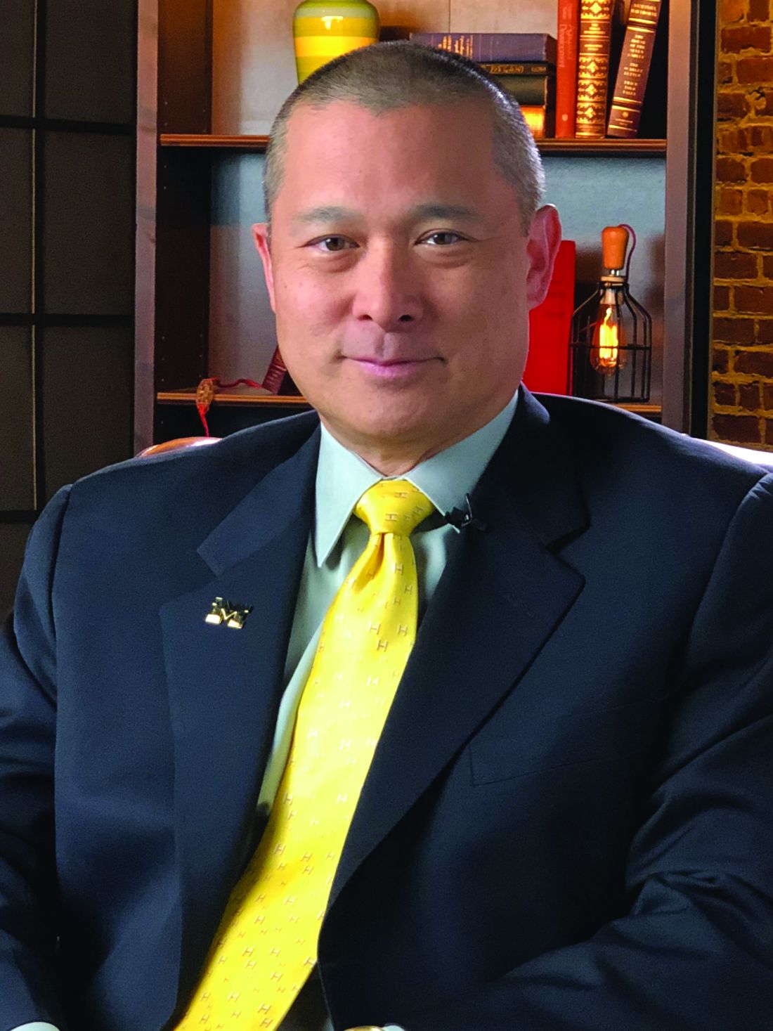
He noted that the low rate of just 50% of recipients of positive FITs going on to receive a colonoscopy is consistent with what is observed in other trials.
“Other data suggests that the rate might even be significantly higher — at 70%-80%, depending upon the population and the test,” Chey told Medscape Medical News.
Reasons for the failure to receive the follow-up testing range from income restrictions (due to the high cost of a colonoscopy, especially if not covered by insurance), education, speaking a foreign language, and other factors, he said.
The relatively high rates of colon cancers detected by FIT in the study, in those with and without a prior polypectomy, along with findings from other studies “should raise questions about whether there might be a role for FIT testing in addition to colonoscopy.” However, much stronger evidence would be needed, Chey noted.
In the meantime, a key issue is “how do we do a better job of making sure that individuals who have a positive FIT test get a colonoscopy,” he said.
“I think a lot of this is going to come down to how it’s done at the primary care level.”
Chey added that in that, and any other setting, “the main message that needs to get out to people who are undergoing stool-based screening is that the stool test is only the first part of the screening process, and if it’s positive, a follow-up colonoscopy must be performed.”
“Otherwise, the stool-based test is of no value.”
Wilson had no disclosures to report. Chey’s disclosures included consulting and/or other relationships with Ardelyx, Atmo, Biomerica, Commonwealth Diagnostics International, Corprata, Dieta, Evinature, Food Marble, Gemelli, Kiwi BioScience, Modify Health, Nestlé, Phathom, Redhill, Salix/Valeant, Takeda, and Vibrant.
A version of this article appeared on Medscape.com .
PHOENIX — Patients with or without polyp removal in an index colonoscopy commonly receive follow-up surveillance with a fecal immunochemical test (FIT), yet many of these patients do not receive a recommended colonoscopy after a positive FIT.
“In this large US study, we found interval FITs are frequently performed in patients with and without prior polypectomy,” said first author Natalie J. Wilson, MD, of the University of Minnesota in Minneapolis, while presenting the findings at the American College of Gastroenterology (ACG) 2025 Annual Scientific Meeting.
“ and colorectal cancer, regardless of polypectomy history,” Wilson said.
Guideline recommendations stress the need for follow-up surveillance with a colonoscopy, particularly in patients who have had a prior polypectomy, because of the higher risk.
Reasons patients may instead turn to FIT may include cost or other factors, she said.
To determine just how often that happens, how having a previous polypectomy affects FIT results, and how adherent patients are to follow up if a FIT result is positive, Wilson and her colleagues evaluated data from nearly 4.8 million individuals in the Veterans Health Administration Corporate Data Warehouse who underwent colonoscopy between 2000 and 2024.
Of the patients, 10.9% were found to have subsequently received interval FIT within 10 years of the index colonoscopy, and of those patients, nearly half (49.9%) had received a polypectomy at the index colonoscopy.
The average time from the colonoscopy/polypectomy to the interval FIT was 5.9 years (5.6 years in the polypectomy group vs 6.2 years in the non-polypectomy group).
Among the FIT screenings, results were positive in 17.2% of post-polypectomy patients and 14.1% of patients with no prior polypectomy, indicating a history of polypectomy to be predictive of a positive interval FIT (odds ratio [OR], 1.12; P < .0001).
Notably, while a follow-up colonoscopy is considered essential following a positive FIT result — and having a previous polypectomy should add further urgency to the matter — the study showed only 50.4% of those who had an earlier polypectomy went on to receive the recommended follow-up colonoscopy after a positive follow-up FIT, and the rate was 49.3% among those who had not received a polypectomy (P = .001).
For those who did receive a follow-up colonoscopy after a positive FIT, the duration of time to receiving the colonoscopy was longer among those who had a prior polypectomy, at 2.9 months compared with 2.5 months in the non-polypectomy group (P < .001).
Colonoscopy results following a positive FIT showed higher rates of detections among patients who had prior polypectomies than among those with no prior polypectomy, including tubular adenomas (54.7% vs 45.8%), tubulovillous adenomas (5.6% vs 4.7%), adenomas with high-grade dysplasia (0.8% vs 0.7%), sessile serrated lesions (3.52% vs 2.4%), advanced colorectal neoplasia (9.2% vs 7.9%), and colorectal cancer (3.3% vs 3.0%).
However, a prior polypectomy was not independently predictive of colorectal cancer (OR, 0.96; P = .65) or advanced colorectal neoplasia (OR, 0.97; P = .57) in the post-colonoscopy interval FIT.
The findings underscore that “positive results carried a high risk of advanced neoplasia or cancer, irrespective of prior polypectomy history,” Wilson said.
Clinicians Must ‘Do a Better Job’
Commenting on the study, William D. Chey, MD, AGAF, chief of the Division of Gastroenterology & Hepatology at the University of Michigan in Ann Arbor, noted that the study “addresses one of the biggest challenges we face as a profession, which is making sure that patients who have a positive stool test get a colonoscopy.”
He noted that the low rate of just 50% of recipients of positive FITs going on to receive a colonoscopy is consistent with what is observed in other trials.
“Other data suggests that the rate might even be significantly higher — at 70%-80%, depending upon the population and the test,” Chey told Medscape Medical News.
Reasons for the failure to receive the follow-up testing range from income restrictions (due to the high cost of a colonoscopy, especially if not covered by insurance), education, speaking a foreign language, and other factors, he said.
The relatively high rates of colon cancers detected by FIT in the study, in those with and without a prior polypectomy, along with findings from other studies “should raise questions about whether there might be a role for FIT testing in addition to colonoscopy.” However, much stronger evidence would be needed, Chey noted.
In the meantime, a key issue is “how do we do a better job of making sure that individuals who have a positive FIT test get a colonoscopy,” he said.
“I think a lot of this is going to come down to how it’s done at the primary care level.”
Chey added that in that, and any other setting, “the main message that needs to get out to people who are undergoing stool-based screening is that the stool test is only the first part of the screening process, and if it’s positive, a follow-up colonoscopy must be performed.”
“Otherwise, the stool-based test is of no value.”
Wilson had no disclosures to report. Chey’s disclosures included consulting and/or other relationships with Ardelyx, Atmo, Biomerica, Commonwealth Diagnostics International, Corprata, Dieta, Evinature, Food Marble, Gemelli, Kiwi BioScience, Modify Health, Nestlé, Phathom, Redhill, Salix/Valeant, Takeda, and Vibrant.
A version of this article appeared on Medscape.com .
FROM ACG 2025
Polypectomy Best Practices Not Routinely Followed in US
, an analysis of more than 1.8 million colonoscopies found.
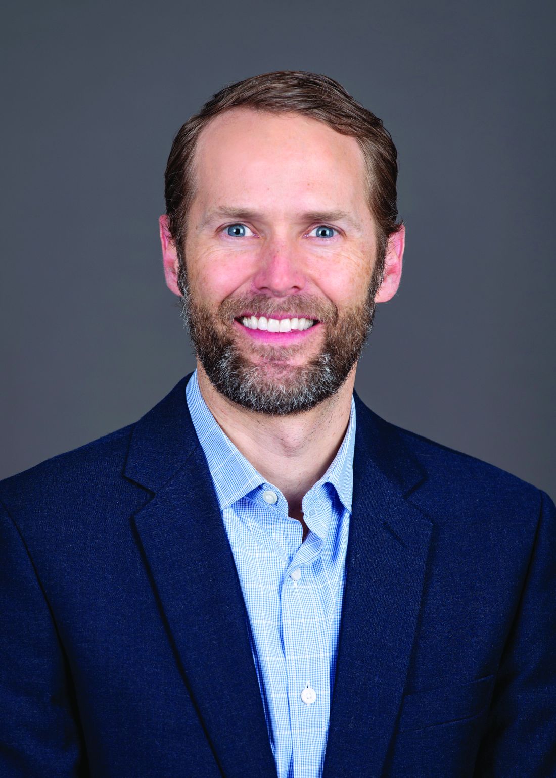
“We expected to find some variations in polypectomy technique, but the results were surprising; overall, cold snare usage was much lower than expected, given that this is the recommended method for removing most small polyps,” Seth Crockett, MD, MPH, AGAF, professor of medicine, Division of Gastroenterology and Hepatology, Oregon Health & Science University, Portland, told GI & Hepatology News.
The study was published in the October issue of The American Journal of Gastroenterology.
Using Gastroenterology Quality Improvement Consortium Registry data, Crockett and colleagues analyzed more than 1.8 million colonoscopies performed by 4601 endoscopists between 2019 and 2022 across 702 sites. All colonoscopies involved removal of polyps < 1 cm; lesions of this size are commonly found in screening colonoscopies, and detection is crucial to early cancer prevention.
The researchers found striking variation in polypectomy technique. Guideline-based cold snare polypectomy (CSP) was used in only 58% of cases (and as a single device in only 51%), whereas cold forceps polypectomy (CFP) accounted for 35% and hot snare polypectomy (HSP) for 11%.
The fact that CSP was used in fewer than 60% of cases represents “an important quality gap,” the authors wrote, adding that the fact that more than 10% of colonoscopies used HSP suggests that “some patients harboring low-risk lesions may be exposed to excess risk related to these practice variations.”
And while recommendations around the use of CFP are more nuanced (based largely on forceps type and polyp size), the “high frequency of CFP also suggests nonadherence to best practices,” they noted.
Gastroenterologists More Apt to Follow Guidance
Polypectomy technique varied by polyp type. CFP was more common in cases where only hyperplastic polyps were removed compared with cases with tubular adenomas (45% vs 30%, respectively). CSP use was highest in cases where only sessile serrated lesions were removed (66%) compared with cases with only tubular adenomas (61%) or hyperplastic polyps (37%).
There was also considerable variation by provider specialty.
Gastroenterologists (compared with non-GI specialists) used HSP less (4% vs 8%) and CSP more (40% vs 34%). Colonoscopies performed with GI fellows were more likely to use CFP (31% vs 21%) and less likely to use HSP (1% vs 5%) compared with colonoscopies without fellows.
“It was somewhat reassuring that colonoscopies performed by gastroenterologists were more likely to adhere to guideline recommendations, which suggests that dedicated endoscopy training is likely an important factor driving high-quality colonoscopy,” Crockett told GI & Hepatology News.
“Unexpectedly,” polypectomy technique also differed dramatically by geographic region, he said. CFP was used more than twice as often in the Northeast (31%) as in the Midwest (14%), whereas CSP was used more frequently in the Midwest (52%) than in the Northeast (32%).
“We suspect that much of the variation is related to differences in training, preferences, habits, and evolution of colonoscopy practice over time,” Crockett said. “More research is needed on the underlying drivers of this variation, and how differences in polypectomy technique impact both the safety and efficacy of colonoscopy to prevent colorectal cancer,” he said.
“As a specialty, we need to continue to work on disseminating guideline recommendations regarding colonoscopy quality, monitoring adherence to evidence-based practices, and working to address gaps in quality where they exist,” he added.
‘Concerning, Surprising, and Disappointing’
David Johnson, MD, professor of medicine and chief of gastroenterology at Eastern Virginia Medical School and Old Dominion University in Norfolk, called the results “concerning, surprising, and disappointing” and not consistent with the most current quality recommendations that advocate cold snare for most polyps less than 1 cm in size.
“Cold snare polypectomy has been shown not only to be more effective but also takes less time to perform, relative to cold biopsy,” said Johnson, who wasn’t involved in the study.
Johnson told GI & Hepatology News, “Inadequate lesion resection and variation in resection quality are major issues for colonoscopy quality. Those who perform colonoscopies need to be up-to-date with evidence-based quality standards — as well as held accountable if [there is] discordant practice — if we are to optimize the cancer prevention benefits of quality colonoscopy.”
Limitations of the current analysis include lack of extensive patient information and inability to further stratify polyps < 1 cm by size.
The study had no commercial funding. Crockett had no disclosures. Johnson disclosed serving as a director, officer, partner, employee, advisor, consultant, or trustee for ISOThrive.
A version of this article appeared on Medscape.com.
, an analysis of more than 1.8 million colonoscopies found.
“We expected to find some variations in polypectomy technique, but the results were surprising; overall, cold snare usage was much lower than expected, given that this is the recommended method for removing most small polyps,” Seth Crockett, MD, MPH, AGAF, professor of medicine, Division of Gastroenterology and Hepatology, Oregon Health & Science University, Portland, told GI & Hepatology News.
The study was published in the October issue of The American Journal of Gastroenterology.
Using Gastroenterology Quality Improvement Consortium Registry data, Crockett and colleagues analyzed more than 1.8 million colonoscopies performed by 4601 endoscopists between 2019 and 2022 across 702 sites. All colonoscopies involved removal of polyps < 1 cm; lesions of this size are commonly found in screening colonoscopies, and detection is crucial to early cancer prevention.
The researchers found striking variation in polypectomy technique. Guideline-based cold snare polypectomy (CSP) was used in only 58% of cases (and as a single device in only 51%), whereas cold forceps polypectomy (CFP) accounted for 35% and hot snare polypectomy (HSP) for 11%.
The fact that CSP was used in fewer than 60% of cases represents “an important quality gap,” the authors wrote, adding that the fact that more than 10% of colonoscopies used HSP suggests that “some patients harboring low-risk lesions may be exposed to excess risk related to these practice variations.”
And while recommendations around the use of CFP are more nuanced (based largely on forceps type and polyp size), the “high frequency of CFP also suggests nonadherence to best practices,” they noted.
Gastroenterologists More Apt to Follow Guidance
Polypectomy technique varied by polyp type. CFP was more common in cases where only hyperplastic polyps were removed compared with cases with tubular adenomas (45% vs 30%, respectively). CSP use was highest in cases where only sessile serrated lesions were removed (66%) compared with cases with only tubular adenomas (61%) or hyperplastic polyps (37%).
There was also considerable variation by provider specialty.
Gastroenterologists (compared with non-GI specialists) used HSP less (4% vs 8%) and CSP more (40% vs 34%). Colonoscopies performed with GI fellows were more likely to use CFP (31% vs 21%) and less likely to use HSP (1% vs 5%) compared with colonoscopies without fellows.
“It was somewhat reassuring that colonoscopies performed by gastroenterologists were more likely to adhere to guideline recommendations, which suggests that dedicated endoscopy training is likely an important factor driving high-quality colonoscopy,” Crockett told GI & Hepatology News.
“Unexpectedly,” polypectomy technique also differed dramatically by geographic region, he said. CFP was used more than twice as often in the Northeast (31%) as in the Midwest (14%), whereas CSP was used more frequently in the Midwest (52%) than in the Northeast (32%).
“We suspect that much of the variation is related to differences in training, preferences, habits, and evolution of colonoscopy practice over time,” Crockett said. “More research is needed on the underlying drivers of this variation, and how differences in polypectomy technique impact both the safety and efficacy of colonoscopy to prevent colorectal cancer,” he said.
“As a specialty, we need to continue to work on disseminating guideline recommendations regarding colonoscopy quality, monitoring adherence to evidence-based practices, and working to address gaps in quality where they exist,” he added.
‘Concerning, Surprising, and Disappointing’
David Johnson, MD, professor of medicine and chief of gastroenterology at Eastern Virginia Medical School and Old Dominion University in Norfolk, called the results “concerning, surprising, and disappointing” and not consistent with the most current quality recommendations that advocate cold snare for most polyps less than 1 cm in size.
“Cold snare polypectomy has been shown not only to be more effective but also takes less time to perform, relative to cold biopsy,” said Johnson, who wasn’t involved in the study.
Johnson told GI & Hepatology News, “Inadequate lesion resection and variation in resection quality are major issues for colonoscopy quality. Those who perform colonoscopies need to be up-to-date with evidence-based quality standards — as well as held accountable if [there is] discordant practice — if we are to optimize the cancer prevention benefits of quality colonoscopy.”
Limitations of the current analysis include lack of extensive patient information and inability to further stratify polyps < 1 cm by size.
The study had no commercial funding. Crockett had no disclosures. Johnson disclosed serving as a director, officer, partner, employee, advisor, consultant, or trustee for ISOThrive.
A version of this article appeared on Medscape.com.
Durvalumab Plus FLOT Ups Survival in Early Upper-GI Cancer
BERLIN — , according to findings presented at the 2025 annual meeting of the European Society for Medical Oncology (ESMO).
Experts said the survival benefit further supports perioperative durvalumab plus FLOT (fluorouracil, leucovorin, oxaliplatin, and docetaxel) as the new standard of care for patients with localized gastric or gastroesophageal adenocarcinoma. Earlier results from the phase 3 MATTERHORN trial, reported at the American Society of Clinical Oncology meeting (ASCO) in June, showed that the addition of durvalumab improved event-free survival compared with FLOT alone.
The findings presented at ESMO show that at 36 months, overall survival was 68.6% among patients who received durvalumab + FLOT vs 61.9% among those given FLOT plus a placebo. After a median of 43 months, the survival advantage in the durvalumab group was statistically significant (hazard ratio [HR], 0.78; 95% CI, 0.63-0.96; P = .021) and “more importantly, clinically meaningful,” said lead investigator Josep Tabernero, MD, PhD, of Vall d’Hebron University Hospital in Barcelona.
The results “strongly support the use of perioperative durvalumab plus chemotherapy with FLOT as a new global standard of care for patients with localized gastric and gastroesophageal adenocarcinoma,” Tabernero said.
Speaking as discussant for the session, Sylvie Lorenzen, MD, PhD, Technische Universität München in Munich, Germany, was enthusiastic that the previously reported trends in MATTERHORN held strong.
“The shape of the curves presented at ASCO was already very positive,” she said. “And now, with a longer follow-up, more events, and a higher overall survival maturity, they reach statistical significance. It looks like the magnitude of the effect increases with longer follow-up, and this is important for our patients.”
The trial randomly assigned 948 patients with resectable gastric or gastroesophageal adenocarcinoma to receive either durvalumab (1500 mg) or placebo every 4 weeks, plus FLOT for 2 cycles before surgery and then again after, followed by durvalumab or placebo every 4 weeks for 10 cycles.
Patients were stratified according to lymph node status, as well as PD-L1 expression (≥ 1% or < 1%, according to the Tumor Area Positivity score.)
The improvement in overall survival with durvalumab was seen regardless of PD-L1 expression, Tabernero said, with the same hazard ratios (0.79) in both the positive and negative subgroups.
However, there was no clear overall survival benefit in certain other subgroups, including women (n = 266; HR, 0.91), those with node-negative disease (n = 277; HR, 1.01), and those with diffuse histology (n = 249; HR, 0.98).
Lorenzen said that clinicians should “pay attention” to those patient subgroups, as they seem to benefit less from the addition of durvalumab. However, she cautioned that the findings were based on small patient numbers and the confidence intervals were wide.
Tabernero also reported additional data on event-free survival (EFS). Overall, the durvalumab/FLOT combination improved EFS among patients with any degree of pathological response and irrespective of lymph node status at surgery.
Regarding nodal staging, which was done in 800 patients, the percentage who achieved negative nodal status was higher in the durvalumab group (58.2%) vs the placebo group (44.8%). However, the improvement in EFS with durvalumab was comparable for node-negative (HR, 0.74) and node-positive (HR, 0.77) patients.
Lorenzen said that overall, the results provide a solid answer to the question, “Is it time to change practice?”
“I think MATTERHORN gives us the largest dataset and answers this question satisfactorily,” she said. Given that overall survival improved regardless of PD-L1 expression, she added, the combination of durvalumab and FLOT should be offered to “all our patient subgroups.”
The study was funded by AstraZeneca. Tabernero made numerous disclosures, including relationships with AstraZeneca, Boehringer Ingelheim, Chugai, and Daichii Sankyo. Lorenzen disclosed financial interests in or serving as an invited speaker for Servier, Lilly, MSD, and BMS.
A version of this article appeared on Medscape.com .
Half of Patients Skip Repeat Stool Tests for CRC Screening
A large real-world study found that
Among those who did repeat the test, the average delay was 3 months before COVID and increased to 5 months during the pandemic, the authors reported in BMJ Public Health.
“Stool tests are relatively easy to complete at home and mailed for testing, and they are inexpensive, but they must be completed annually. In contrast, colonoscopies are more invasive and require more time away from work but only need to be repeated every 5-10 years,” Staci J Wendt, PhD, director, health research accelerator, Providence Research Network, Providence, Rhode Island, told GI & Hepatology News.
In the end, “the best colorectal cancer screening test is the one that gets done,” Wendt said.
“This is why we stress the importance of patients and their doctor having these discussions together and deciding which screening is the most preferred method for the individual patient,” she added.
Stool Tests Gaining Traction
Adults are increasingly turning to at-home stool tests for CRC screening — a trend that accelerated during the pandemic. Yet, there is limited data on whether patients undergo repeat stool tests following initial negative test results.
Wendt and her colleagues documented rates of repeat preventative stool tests by analyzing electronic medical records from Providence St Joseph Health, a large health system with 51 hospitals and over 1000 clinics across seven western US states.
They divided their analysis into two periods based on the onset of the pandemic. The pre-COVID onset period spanned January 2018 to February 2020 and the post-COVID period spanned March 2020 to February 2022.
“The pandemic is a salient time to conduct this study because it resulted in a dramatic decrease in colonoscopies, which were partially replaced by stool tests. This partial replacement of colonoscopies by stool tests has led other studies to conclude that stool tests mitigated gaps in CRC screening during the pandemic. But gaps may persist if patients do not undergo repeat testing,” the study team explained.
Their sample included 403,085 patients. Among those with an initial negative stool test, the share who obtained a timely repeat screening ranged from 38% to 49% across the study years, confirming that “most patients do not undergo the recommended repeat screening after their initial stool test,” the researchers said.
Among adults who do a repeat test, delays were common. The average lag to the follow-up test was 3months on average, increasing to about 5 months amid COVID — almost half as long as the preventative screening period of stool tests (12 months).
“These gaps could delay detection of CRC and subsequent treatment, potentially resulting in higher mortality. These gaps are particularly important as more and more patients use stool tests instead of colonoscopes for CRC screening,” the researchers wrote.
Screening patterns shifted markedly during the pandemic.
Not surprisingly, the volume of colonoscopies declined substantially after the onset of the pandemic and stayed low through the study’s end. In contrast, the volume of at-home stool tests was increasing before the pandemic and accelerated during the pandemic.
“Given this increase in stool tests, it will be increasingly important to focus on improving long-term adherence to screening through outreach, policies and programs,” the researchers said.
A Multilevel Approach
Wendt said health systems that are incorporating proactive measures like sending stool kits to patients who are eligible for screening, should ensure that these screening kits and information are sent annually and that it is stressed that the screening must happen every year.
Reached for comment, Aasma Shaukat, MD, MPH, AGAF, director of outcomes research, Division of Gastroenterology and Hepatology, NYU Langone Health, New York City, who wasn’t involved in the study, said the poor adherence to repeat stool tests for CRC screening seen in this study is “not surprising.”

“We know that adherence goes down with each consecutive screening round and what is really needed is an organized program to keep the level of adherence up,” Shaukat told GI & Hepatology News.
Shaukat agreed that boosting adherence to stool tests requires a “multilevel approach.”
She cited the success of the CRC screening program implemented across Kaiser Permanente Northern California. The program includes proactive and targeted outreach to members who are overdue for screening and mailed fecal immunochemical test kits for at-home use.
As reported previously by GI & Hepatology News, the program has made a huge difference in CRC incidence, deaths, and racial disparities.
The program has doubled the proportion of people up to date with screening. And, within about 10 years, cancer rates were cut by a third, deaths were halved and largely eliminated long-standing differences by race and ethnicity.
The study had no commercial funding. Wendt and Shaukat declared having no relevant disclosures.
A version of this article appeared on Medscape.com.
A large real-world study found that
Among those who did repeat the test, the average delay was 3 months before COVID and increased to 5 months during the pandemic, the authors reported in BMJ Public Health.
“Stool tests are relatively easy to complete at home and mailed for testing, and they are inexpensive, but they must be completed annually. In contrast, colonoscopies are more invasive and require more time away from work but only need to be repeated every 5-10 years,” Staci J Wendt, PhD, director, health research accelerator, Providence Research Network, Providence, Rhode Island, told GI & Hepatology News.
In the end, “the best colorectal cancer screening test is the one that gets done,” Wendt said.
“This is why we stress the importance of patients and their doctor having these discussions together and deciding which screening is the most preferred method for the individual patient,” she added.
Stool Tests Gaining Traction
Adults are increasingly turning to at-home stool tests for CRC screening — a trend that accelerated during the pandemic. Yet, there is limited data on whether patients undergo repeat stool tests following initial negative test results.
Wendt and her colleagues documented rates of repeat preventative stool tests by analyzing electronic medical records from Providence St Joseph Health, a large health system with 51 hospitals and over 1000 clinics across seven western US states.
They divided their analysis into two periods based on the onset of the pandemic. The pre-COVID onset period spanned January 2018 to February 2020 and the post-COVID period spanned March 2020 to February 2022.
“The pandemic is a salient time to conduct this study because it resulted in a dramatic decrease in colonoscopies, which were partially replaced by stool tests. This partial replacement of colonoscopies by stool tests has led other studies to conclude that stool tests mitigated gaps in CRC screening during the pandemic. But gaps may persist if patients do not undergo repeat testing,” the study team explained.
Their sample included 403,085 patients. Among those with an initial negative stool test, the share who obtained a timely repeat screening ranged from 38% to 49% across the study years, confirming that “most patients do not undergo the recommended repeat screening after their initial stool test,” the researchers said.
Among adults who do a repeat test, delays were common. The average lag to the follow-up test was 3months on average, increasing to about 5 months amid COVID — almost half as long as the preventative screening period of stool tests (12 months).
“These gaps could delay detection of CRC and subsequent treatment, potentially resulting in higher mortality. These gaps are particularly important as more and more patients use stool tests instead of colonoscopes for CRC screening,” the researchers wrote.
Screening patterns shifted markedly during the pandemic.
Not surprisingly, the volume of colonoscopies declined substantially after the onset of the pandemic and stayed low through the study’s end. In contrast, the volume of at-home stool tests was increasing before the pandemic and accelerated during the pandemic.
“Given this increase in stool tests, it will be increasingly important to focus on improving long-term adherence to screening through outreach, policies and programs,” the researchers said.
A Multilevel Approach
Wendt said health systems that are incorporating proactive measures like sending stool kits to patients who are eligible for screening, should ensure that these screening kits and information are sent annually and that it is stressed that the screening must happen every year.
Reached for comment, Aasma Shaukat, MD, MPH, AGAF, director of outcomes research, Division of Gastroenterology and Hepatology, NYU Langone Health, New York City, who wasn’t involved in the study, said the poor adherence to repeat stool tests for CRC screening seen in this study is “not surprising.”
“We know that adherence goes down with each consecutive screening round and what is really needed is an organized program to keep the level of adherence up,” Shaukat told GI & Hepatology News.
Shaukat agreed that boosting adherence to stool tests requires a “multilevel approach.”
She cited the success of the CRC screening program implemented across Kaiser Permanente Northern California. The program includes proactive and targeted outreach to members who are overdue for screening and mailed fecal immunochemical test kits for at-home use.
As reported previously by GI & Hepatology News, the program has made a huge difference in CRC incidence, deaths, and racial disparities.
The program has doubled the proportion of people up to date with screening. And, within about 10 years, cancer rates were cut by a third, deaths were halved and largely eliminated long-standing differences by race and ethnicity.
The study had no commercial funding. Wendt and Shaukat declared having no relevant disclosures.
A version of this article appeared on Medscape.com.